By: Mark Melin, Heather Hettrick, Monika Gloviczki, Stanley G. Rockson, Leonhard Möckl, Eno Ebong, Frank Aviles Jr.
Time and gravity are perpetual invisible forces impacting human physiology from conception until death. Time is measured by the gravity-induced swing of a grandfather clock’s pendulum, the red digits of an atomic clock, the composer’s notes upon a sheet of piano music, the graying of hair, and the presence of grandchildren of the next generation.
The constant downward acceleration of gravity is measured by a gravimeter, variable in strength depending upon the height and location of the local environment on earth, from the seabed floor to the heights of Mount Everest. Gravity’s impact upon human structure and function is noted in the time progressive wrinkling and “sagging” of tissues, the muscle tone defining and bone strength building of resistive weight training, the augmentation of jugular vein and facial lymphatic drainage, and the compromise of microcirculatory perfusion due to significant interstitial edema in the lower extremities of patients with advanced venous hypertension (lymphedema of venous etiology/ phlebolymphedema) and stage III lymphedema, resulting in dermal ulcer development.
The goal of this overview is to introduce the factors that impact the health of the international astronaut core during spaceflight (weightlessness or decreased gravity environment) and the possible translation of this extensive expanding data set to better understand the pathophysiology of our patients in “1G” wound clinics and hospitals due to the extreme disease states that have risen to epidemic levels in diabetes mellitus, obesity, and venous insufficiency, all now known to be concomitant with lymphedema.
Gravity and Weightlessness
Ground-based analogs for simulation of the impact of weightlessness upon human physiology, providing for the performance of extensive research opportunities, exist in several forms, including days/weeks of strict 6-degree head-down tilt bed rest (HDTBR), dry immersion, and brief periods (20–30 seconds) of weightlessness during parabolic flights.1,2 The elevated CO2 levels of the International Space Station (ISS) environment are related to human exhalation and to limits related to CO2 “scrubbing” on the ISS. The average earth atmospheric carbon dioxide level is 410 ppm. The ISS maximum 24-hour allowable CO2 levels are ten times higher at 5250 ppm (the breathing environment on the ISS is 14.7 psi with normal oxygen and nitrogen levels).
Astronauts may also be exposed to pockets of even higher CO2 levels in areas of low ventilation, given that there is no natural convection in microgravity. Elevated CO2 levels have also been simulated in head-down tilt studies, specifically for Spaceflight Associated Neuro-ocular Syndrome (SANS) research.3,4
Studies on astronauts and cosmonauts in true weightlessness have been performed in all spaceflight eras, from Mercury to Apollo to the ISS, including the 25-month- long NASA twins study, that compared monozygotic twin astronauts through “extensive integrated longitudinal, multidimensional measures of physiological, telomeric, transcriptomic, epigenetic, proteomic, metabolomic, immune, microbiomic, cardiovascular, vision-related and cognitive data,” as one brother spent 340 days in space on the ISS, and his twin remained earthbound.5,6
We encourage you to read this tour de force study produced by 84 world-class researchers across 12 universities, NASA, the European Space Agency (ESA) and additional resources. Brinda Rhana, PhD, UC San Diego School of Medicine, and a member of the team responsible for monitoring cardiac changes and adaptations, says that the study has broad findings that “demonstrated on the molecular level the resilience and robustness of how a human body can adapt to a multitude of changes induced by the space- flight environment, such as microgravity (weightlessness), radiation, circadian disruption, elevated CO2, isolation from friends and family and dietary limitations.”
Gravity is the weakest of the four fundamental forces in nature (which also include electromagnetic force and strong and weak nuclear forces), but it dominates over large distances, is only attractive, and can never be screened from its interaction between two bodies. In “1G”, 70% of body fluids reside below the level of the heart. The lymphatic system has the capacity and capability to transport fluid against gravity as well as tissue pressure gradients via lymphangion contractility, leg muscle contraction, and respiratory and chest wall function, thus creating a “suction” effect for pumping lymphatic fluid within the subatmospheric pressure tissue distribution zones (the Guyton principle).7
In the weightlessness of space and the loss of the 1G head-to-foot hydrostatic pressure gradient, astronauts experience a dramatic fluid redistribution of about two liters from the legs to the head, neck, and upper torso within the first 24–48 hours of flight. In the first two weeks, they experience a 10–17% plasma volume reduction, increased cardiac output and stroke volume (Doppler technique on the ISS, 23–25% and 19–21%, respectively), decreased systemic vascular resistance (SVR) of 14–39%, the phenomenon of neocytolysis resulting in young red blood cell culling and a relative decline in red blood cell total mass, increased oxidative stress and inflammation with the potential to impact arterial function, among other cardiovascular system and body system adaptations.8-18
Adaptations are particularly evident in the venous system, with decreased calf venous volume, increased venous compliance, and altered venous filling and emptying functions noted on-air plethysmography on the ISS.19 To determine if six months in microgravity results in significant changes in the major central and peripheral veins, indicating a redistribution of venous blood flow, ten astronauts were assessed by echography on the ISS for the measurement of vessel cross-sectional area of the jugular vein, portal vein, femoral vein, tibial vein, and gastrocnemius vein. The inflight exams were conducted by astronauts using a volume capture method in which images collected were processed to produce a 3D reconstruction of the vessel that was later analyzed by a trained sonographer. Measurements were conducted pre-flight, at the beginning of the flight (day 15), near the end of the flight (4–5.5 months), and post-flight.
The increase in jugular, portal, and femoral vein cross-sectional area during spaceflight confirmed that there was venous blood pooling in the cephalic, splanchnic and pelvic regions. In contrast, the tibial veins were found to be decreased with spaceflight, related to the cephalad fluid shifts. All veins returned to baseline conditions four days after returning to earth.20
Unbalanced cerebral arterial inflow and decreased venous outflow have the potential to create a relative increased intracranial pressure (ICP) and intracranial/extracranial parenchymal and soft tissue edema based on a modified understanding of the Monro-Kellie doctrine.21 IJV distension has been demonstrated with ultrasound in a prospective cohort study of 11 astronauts on the ISS during long-duration spaceflight missions. The mean IJV area increased from 9.8 mm2 in the preflight seated position to 70.3 mm2 during spaceflight, with an associated mean IJV pressure increase from 5.1 mm Hg to 21.1 mm Hg. One crew member was noted to have an asymptomatic occlusive IJV thrombus and retrospectively, a second potential partial IJV thrombus was identified in a second crew member.22
Treatment of the IJV deep venous thrombus (DVT) included enoxaparin (onboard the ISS’s medicine cabinet) and eventual delivery of oral apixaban, protamine, and prothrombin complex concentrate to the space station with a supply spacecraft 42 days later. Therapy was transitioned to apixaban with ongoing sonographic surveillance and ground-based telemedicine multidisciplinary management until deorbit and landing. Apixaban was stopped four days before landing. A point-of-care ultra- sound upon spacecraft landing revealed spontaneous flow in the IJV with residual thrombus flattened to the vessel wall. A subsequent thrombophilic work up was unremarkable.23
Lymphatic Research
Lymphatic research has experienced a renaissance in the past decade with the recognition and increasing knowledge of the dynamic nature of the endothelial glycocalyx and the resulting modification of the classical Starling principle to reflect the true critical nature of lymphatic integrative function with arterial, venous, integument, and immune health.
The recognition of modified Starling forces now confirms that the majority of interstitial fluid resulting from arterial perfusion re-enters the central venous system via the vast lymphatic network and not via venules.24–29 Despite new significant milestones in research and clinical application, lymphatic education remains “paradoxically and unnecessarily ignored” at the medical school and residency levels.30
Lymphatic research in space and astronaut health may paradoxically elevate the subject on terra firma. Chronic venous hypertension (CVI) and all venous ulcerations have associated glycocalyx thinning or shed- ding.31 The associated lymphedema of venous etiology/ phlebolymphedema is common though vastly under-recognized and undertreated component and has a significant economic impact.32-34 There remain substantial gaps in the understanding of the role of topical treatment of venous leg ulcers and associated lymphedema of venous etiology.35 The potential for patient care improvement (as translated to earth-bound medical care, a “spinoff”) may be further elevated with the continued focus on astronaut health, countermeasure development, the near-term space tourism era, and the ambitious near-term goals of moon habitation and Mars human exploration.36
Lymphatic research in low Earth orbit (LEO) is in the initial phases of development, though has had vital forward-thinking and visionary investigator-led early research. Published data have used mice and rats in studies involving tail suspension and other altered gravity models to evaluate fluid shifts as countermeasures have undergone development.37-39 Specially designed satellites with mouse habitats have been used for research. Space shuttle era research has been used to explore the potential translational aspects of immune function in the context of astronaut stress, environment, and activities. Plasma cytokines and recently published data on astronaut saliva cytokine measurements confirm an in-vivo hormonal dysregulation of immunity, appearing pro-inflammatory and persisting during long-duration orbital spaceflight.40 As astronauts increase exploration limits, these biomarkers will be important for monitoring the effectiveness of biomedical countermeasures including pharmacology and adjunctive micronutrient use, exercise for bone and muscle health, and radiation shielding. Precision and general environmental counter-measures are now being developed for planned long-term deep-space missions, including pharmacology, environmental filters, radiation shielding, and immune stabilizing nutritional components.41
Rooney et al. published a 2019 paper regarding her- pes virus reactivation in astronauts during spaceflight and applications for patients on earth. Activation of the hypothalamic-pituitary-adrenal (HPA) and sympathetic-ad- renal-medullary (SAM) axes during spaceflight occurs as measured by increased levels of stress hormones, including cortisol, dehydroepiandrosterone, epinephrine, and norepinephrine. These changes, in addition to decreased cell-mediated immunity, contribute to the reactivation of latent herpes viruses in astronauts. These findings coincide with immune system dysregulation observed in astronauts from both short-duration space shuttle flights and long-duration ISS missions. Developing spaceflight countermeasures to prevent viral reactivation is essential for crew safety and comfort.42
The opportunity to further the understanding of and to develop restorative countermeasures for lymphangion contractility and overall lymphatic function and associated immune function in the extreme environment of space carries the high potential to improve our understanding and treatment of patients that we care for in 1G clinics, who have the extremes of lymphedema that negatively affect the quality of life, outcomes, and economics.
Spaceflight Associated Neuro-ocular Syndrome (SANS)
A now well-recognized potential impact on astronaut vision is Spaceflight Associated Neuro-ocular Syndrome (SANS).43, 44 The working definition of SANS is the presence of any of the following findings postflight (compared to preflight): unilateral or bilateral optic disc edema (variable Frisén grades), globe flattening (as defined qualitatively on imaging), choroidal and retinal folds, hyperopic refractive error shifts (>0.75 Diopters), or focal areas of retinal ischemia (i.e., cotton wool spots). These changes have been documented and correlated with the clinical findings of SANS on ocular (e.g., optical coherence tomography [OCT]), orbital (e.g., ultrasonography and magnetic resonance im- aging [MRI]), and cranial MRI. Visual acuity, Amsler grid, ophthalmoscopy, tonometry, fundus photography, orbital ultrasound, and OCT are available on the ISS and have been essential in documenting the development of the in-flight changes of SANS.
Several non-exclusive theories have been proposed, with overlapping validated data that likely contributes to a multi-hit theory.
- The changes may result from a rise in intracranial pressure (ICP) from cephalad fluid shifts during long-duration space flight
- Compartmentalization of cerebral spinal fluid (CSF) within the orbital optic nerve sheath
- Variations in the genetics of enzymes of the
1-carbon metabolic pathways (single nucleotide polymorphisms [SNPs], e.g. Methylenetetrahydrofolate Reductase [MTHFR] C677T, among many other potential abnormalities such as MTRR A66G and SHMT-1 C1420T)42 leading to mildly elevated homocysteine and insufficiency of B vitamins (B12, B6, folate), uncoupling of endothelial nitric oxide synthetase (eNOS) with decreased endothelial nitric oxide (eNO) production and depleted reserves of antioxidant precursors - Altered testosterone and insulin regulation, resulting in endothelial dysfunction
- Elevated ISS environmental CO2 levels
- Increased radiation exposure, specifically when traveling beyond the Van Allen belt, located 400 to 36,000 miles from the earth’s surface.
Intracranial findings on MRI have also been identified, including moderate pituitary dome concavity, upward shifting of the brain, narrowing of CSF spaces, and a mild increase in ventricle size.45–51
Of particular interest is the astronaut genetic variability component and overlap with patients having venous pathology and recognized MTHFR abnormalities among other single nucleotide polymorphism (SNP) changes, resulting in alterations and inefficiencies of metabolism in the 1-carbon pathways of cellular metabolism. MTHFR abnormalities and venous pathology are a well-established association in peer-reviewed literature.52–55 A recent report identified eNOS polymorphisms to be a risk factor for venous thromboembolism (VTE) in the Asian population, though the pathogenesis of VTE is complex and a single eNOS polymorphism is unlikely to significantly impact its occurrence. Therefore, to better explain the potential correlation between some gene polymorphisms and VTE, further haplotype analysis should be carried out to explore the potential gene interaction.56
MTHFR abnormalities exist in 20–40% of the population at large and were reported to have a 52% prevalence in 88 patients assessed in a single-center wound clinic review (personal communication, July 2021, unpublished data). Countermeasure development of appropriate precision and personalized dosing of vitamins B12, B6, and folate, based on recognized genetic polymorphisms, has the potential to improve endothelial function and recoupling of eNOS in those individuals with MTHFR and other SNP abnormalities and could be developed and standardized as an adjunctive micronutrient therapeutic regime for patients with difficult to heal VLUs, in addition to other recognized effective nutraceuticals such as micronized purified flavonoid fractions (MPFF), vitamin D, antioxidant vitamin C, pentoxifylline and sulodexide (a mixture of 80% fast-moving heparin and 20% of dermatan sulfate).57–63
Gravitational influence has been demonstrated on intraocular pressure, with HDTBR increasing episcleral venous pressure and intraocular pressure by 4–7 mmHg, also demonstrated on flight day one of space shuttle missions with subsequent normalization by flight day four. Both venoconstrictive thigh cuffs and lower-body negative pressure (LBNP) chamber/suits have been tested and trialed as countermeasures to translocate fluid away from the head through the venous system, decreasing intraocular pressure. In addition, LBNP has been shown to increase ground reactive forces, potentially beneficial for maintaining bone and muscle mass by producing gravity-like loads.64, 65
One high-value avenue for research opportunities is determining the impact of venoconstrictive thigh cuffs and LBNP upon the dermal lymphatic ventral medial bundles in ground-based space analogue models, as this may significantly contribute to countermeasure development. Understanding lymphagion contractility response and lymphatic ventral medial bundle flow patterns before, during and after cuff use in HDT studies and their correlation to the findings of decreased ocular pressure, ICP (indirect measurements of transcranial venous sinus Doppler given jugular venous distension and stasis in flow patterns with potential intracranial venous effect) and optic nerve sheath diameters with ultrasound 66, 67 and/or MRI, may have a direct bearing on furthering the understanding of SANS contributing factors relative to intracranial and extracranial lymphatic and venous contributions.
Post-Flight Astronaut Orthostatic Intolerance
Astronauts on the ISS spend an average of two hours per day performing resistive exercises and conditioning on highly modified treadmills to maintain cardiovascular conditioning, muscle mass, and bone strength. Age, pre-flight physiologic status, individualized response to training, nutritional intake, cardiovascular adaptability and genetics may all play a role in astronaut physical status during spaceflight and response to re-entering a 1G environment upon landing, creating a recognized need to further evaluate ground-based analogues.68
In 2015, Lee et al. reported that 60–80% of astronauts experienced orthostatic intolerance during 10 minutes of 80° head-up tilt conducted in the controlled conditions of the laboratory 4–6 hours after landing from long-duration spaceflight.69 The objective of a published 2020 follow-up study 70 was to quantify the effectiveness of the gradient compression garment (GCG) immediately post-landing and during the first day of recovery. End of mission fluid loading consisted of consuming 18–20 ml/kg body weight of sodium chloride-water solution or equivalent dry salt with water with 3–4 meals in the last 12–20 hours before landing.
In collaboration with the manufacturer of JOBST medical compression garments (Essity, Stockholm, Sweden), the GCG was developed as an elastic three-piece garment consisting of two thigh-high stockings and shorts that extend to the bottom of the rib cage that provide a continuous gradient of compression from the feet to the top of the garment. Compression is 55 mm Hg at the ankle and gradually decreases along the leg to 35 mm Hg at the knee and 18 mm Hg at the top of the thigh, further reducing to about 16 mm Hg compression over the abdomen.
The GCGs were constructed for each subject individually. After extraction from the capsule at the landing zone in Russia, astronauts were carried to the medical tent for a brief physical examination by their flight surgeon, then donned the GCG with the assistance of trained operators.
Testing occurred as soon as possible after landing, either in the medical tent at the Soyuz landing site or after transport by helicopter to airports in Kazakhstan. No intravenous fluids (IVF) were administered prior to testing at the landing site, but often astronauts received at least 1 L of IVF during transport by helicopter to the airport. On the day of return, six of the seven astronauts participated in testing in the tent. Subsequent testing occurred either in Germany, Norway or Scotland at the refueling stop during travel back to Houston and again at Johnson Space Center.
The stand test began with the astronaut prone on a mat. A manual blood pressure measurement was obtained by about one minute and 45 seconds of rest, and then the astronaut stood as quickly as possible when a command to stand was issued at two minutes. Astronauts stood facing forward for three and a half minutes. At the completion of the stand test, the test operator asked the astronaut to report their perception of motion sickness, ranging from one (no symptoms) to 20 (nausea to the point of vomiting). Results demonstrated that the use of GCGs throughout the first 24 hours after returning from long-duration spaceflight provided effective protection from the development of orthostatic intolerance during a brief stand test.70
Given that spaceflight decondition- ing could be impactful on astronaut activities when arriving and living in conditions of 17% and 39% of earth’s gravity on the moon and Mars, respectively, physiologic adaptations in the form of devices (passive and/or dynamic compression), daily exercise regimens, hydration, micronutrients to maximize micro and macro-circulation, and element-protecting/ functionally enabling spacesuits will be critical components in the new checklists created to maximize astronaut productivity and safety. They will also be critical to managing the potential for orthostatic hypotension that may occur on non-earth surfaces. Gravity’s impact on human physiology will persist at all limits of human exploration.
Conclusion
Astronaut George Zamka, of Polish ancestry, was the February 2010 STS-130 Space Shuttle Endeavour commander, responsible for delivery of the new ISS Tranquility node module and the seven-windowed cupola, a new location for astronauts to experience the “overview effect” described in Frank White’s 1987 classic book of the impact of space exploration upon the heart and soul of astronauts. Com- mander Zamka described it in this manner: “The world from orbit is experienced, not just seen, like a favorite piece of music. I had the sense that this was the view that poets and artists had imagined for centuries.” 71
Paying honor to his fellow countryman, Zamka returned to earth, intent on expressing his experience in the form of Frédéric Chopin’s masterpieces of musical poetry intertwined with the “earthscape” indelibly etched into his clarity of vision and the yearning of his soul to share the moments of his personal overview effect. The result was Chopin: The Space Concert. This is a masterpiece documentary iv of beauty and grace, complete with spectacular NASA photos and images, accompanied by Chopin’s music as played through the instruments of the Sinfonia Viva Orchestra. The magical inspiration of Chopin: The Space Concert has also been immortalized on a timeless canvas by Dr. Monika Gloviczki (Figure 1).
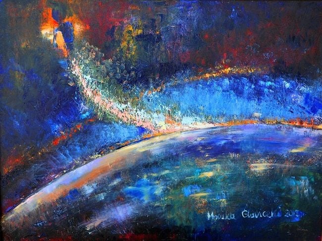
Figure 1: Chopin in the Space by Monika Gloviczki. Oil on canvas, 30 x 40 inches.
Time, one of our greatest allies and treasures, is moving forward. The opportunity is now at hand to advance the science and foundational education of venous and lymphatic physiology, point-of-care clinical application, and the art and science of countermeasure development and treatments. This opportunity can be a derivative, a spinoff, of exploring groundbreaking human venous and lymphatic physiology and adaptability within the extreme environments of weightlessness in LEO, near-term moon habitation, and Mars exploration.
The next frontier is the vertical ascent to travel along the perpetual horizon at 5 miles/second, using multiple validated countermeasures to preserve nominal human physiology in weightlessness. These spinoffs can alter and improve medical school and resident curriculum in the richness and critical components of lymphatic function to human health. They can standardize and normalize the wide spectrum acceptance of venous and lymphedema therapeutics and interventions (payor coverage of quality gradient compression, enhanced coverage of certified lymphedema therapist [CLT] treatment and management, micronized purified flavonoid fraction [MPFF]/ nutraceuticals and adjunctive micronutrients, incorporation of genetics and polygenic assessment to prospectively decrease risk, and standardize the routine use of quality of life-improving advanced lymphedema and arterial pumps), encourage the passage of the Lymphedema Treatment Act and provision for educational assistance to all of our colleagues at all levels of the education scale, and give the tools and instruments to improve and enhance the overall recognition, identification, and care of patients with lymphedema (and lipedema).
Time and gravity are constantly present, as should we be in this exciting, emerging era of venous and lymphatic medicine and surgery. These advances will occur through the sacrifices of the astronaut core from the past, present, and future, upon the spaceships Mercury, Gemini, Apollo, Skylab, Mir, the International Space Station, Space Dragon, Starliner, New Shepard, Unity, and the forthcoming Gateway and Artemis spaceflight systems, and the women and men who perpetually serve and support these missions from their ground-based stations. We applaud, encourage, and congratulate the current basic science and clinical researchers of the collective international research and medical societies in their efforts to advance the field of lymphatic, venous, arterial, and glycocalyx physiology understanding and therapeutic interventions, as benchtop to bedside translation accelerates and results in successful applications.
Godspeed to all as we take up this challenge to carry the baton of advancement and achievement, to hand off to the next generation a medical chapter and text vastly expanded in the understanding of venous and lymphatic physiology
in weightlessness and neogravity. As we expand the field of environmental extremes exploration, personalized nominal function in space travel will vastly improve compassionate care delivery for the patients we serve daily.
References:
1. Pandiarajan M, Hargens AR. “Ground-based Analogs for Spaceflight.” Frontiers in Physiology 2020;11:article 716. https://doi.org/10.3389/ fphys.2020.00716.
2. Cromwell RL, Scott JM, Yarbough PO, et al. “Overview of the NASA
70-day bed rest study.” Med Sci Sports Exerc. 2018;50(9): 1909–1919.
3. Laurie SS, Vizzeri G, Taibbi G, et al. “Effects of short-term mild hypercapnia during head-down tilt on intracranial pressure and ocular structures in healthy human subjects.” Physiol Rep, 5 (11), 2017, e13302. https://doi.org/10.14814/phy2.13302.
4. McGregor HR, Lee JK, Mulder ER, et al. “Ophthalmic Changes in a Spaceflight Analog Are Associated with Brain Functional Reorganization.” Hum Brain Mapp. 2021;1–17. https://doi.org/10.1002/hbm.25546.
5. Berry CA. “Summary of medical experience in the Apollo 7 through 11 manned spaceflights.” Aerospace Medicine 1970, May 500–519.
6. Garrett-Bakelman FE, Darshi M, Green SJ, et al. “The NASA Twins Study: a multidimensional analysis of a year-long human spaceflight.” Science 2019;364, eaau8650.
7. Jamalian S, Jafarnejad M, Zawieja SD, et al. “Demonstration and analysis of the suction effect for pumping lymph fluid from tissue beds at subatmospheric pressure.” Scientific Reports 2017 7:12080. https://doi. org/10.1038/s41598-017-11599-x.
8. Hughson RL, Robertson AD, Shoemaker JK, et al. Increased postflight carotid artery stiffness and inflight insulin resistance resulting from 6 months spaceflight in male and female astronauts. Am J Physiol Heart Circ Physiol 2016 310: H628-H638. Doi:10.1152/ajpheart.00802.2015.
9. Lee SMC, Ribeiro LC, Martin DS, et al. Arterial structure and function during and after long-duration spaceflight. J Appl Physiol 2020;129:108-123.
10. White R. “Weightlessness and the Human Body.” Scientific American, September 1998:59–63.
11. Demontis GC, Germani MM, Caiani EG, Barravecchia I, et al. “Human pathophysiological adaptations to the space environment.” Front Physiol, 02 August 2017. https://doi.org/10.3389/fphys.2017.00547.
12. Norsk, P. “Adaptation of the cardiovascular system to weightlessness: Surprises, paradoxes and implications for deep space missions.” Acta Physiologica 2020;228:e13434. https://doi.org/10.1111/alpha.13434.
13. Norsk P, Asmar A, Damgaard M, Christensen NJ. “Fluid shifts, vasodilation and ambulatory blood pressure reduction during long-duration spaceflight.” J Physiol 2015;593(3):573–584.
14. Diedrich A, Paranjape SY, Robertson D. “Plasma and blood volume in space.” Am J Med Sci 2007;334(1):80-85.
15. Leach CS, Alfrey CP, Suki W, et al. “Regulation of body fluid compartments during short-term spaceflight.” J Appl Physiol 1996;81(1): 105–116.
16. Culliton K, Louati H, Laneuville O, et al. “Six degrees head-down tilt bed rest caused low-grade hemolysis: a prospective randomized clinical trial.” npj Microgravity (2021) 7:4. https://doi.org/10.1038/s41526-021-00132-0.
17. Risso A, Ciana A, Achilli C, et al.” Neocytolysis: none, one or many? A reprisal and future perspectives.” Frontiers in Physiology 2014 5: article 54. https://doi.org/10.3389/fphys.2014.00054.
18. Kunz H, Quiriate H, Simpson RJ, et al. “Alterations in hematologic indices during long-duration spaceflight.” Hematology 2017;17(2): 1–8. https://doi.org/10.1186/s12878-017-0083-y.
19. Fortrat JO, de Holanda A, Zuj K, et al. “Altered venous function during long-duration spaceflights.” Front Physiol 2017;8: article 694. https://doi.org/10.3389/fphys.2017.00694
20. Arbeille P, Provost R, Zuj K, Vincent N. “Measurements of jugular, portal, femoral, and calf vein cross-sectional area for the assessment of venous blood redistribution with long-duration spaceflight (Vessel Imaging Experiment).” Eur J Appl Physiol (2015) 115:2099–2106. https://doi. org/10.1007/s00421-015-3189-6.
21. Wilson MH. “Monro-Kellie 2.0: The dynamic vascular and venous pathophysiological components of intracranial pressure.” Journal of Cerebral Blood Flow & Metabolism 2016; Vol. 36(8): 1338–1350.
22. Marshall-Goebel K, Laurie SS, Alferova IV, et al. “Assessment of jugular venous blood flow stasis and thrombosis during spaceflight.” JAMA Network Open 2019;2(11):e1915011. https://doi.org/10.1001/jamanetworkopen.2019.15011.
23. Auñón-Chancellor SM, Pattarini, JM, Moll, S, Sargsyan, A. “Venous Thrombosis during Spaceflight.” NEJM 2020. 382;1:89–90.
24. Möckl L. “The Emerging Role of the Mammalian Glycocalyx in Functional Membrane Organization and Immune System Regulation.” Front Cell Dev Biol., 15 April 2020. https://doi.org/10.3389/fcell.2020.00253.
25. Mitra R, O’Neil GL, Harding IC, Cheng MJ, Mensah SA, Ebong EE. “Gly-cocalyx in Atherosclerosis-Relevant Endothelium Function and as a Therapeutic Target.” Curr Atheroscler Rep (2017) 19: 63. https://doi. org/10.1007/s11883-017-0691-9.
26. Fu BM, Tarbell JM. “Mechano-sensing and transduction by endothelial surface glycocalyx: composition, structure, and function.” Wiley Interdiscip Rev Syst Biol Med. 2013; 5(3): 381–390. https://doi.org/10.1002/wsbm.1211.
27. Michel CC. “Starling: The formulation of his hypothesis of microvascular fluid exchange and its significance after 100 years.” Experimental Physiology (1997), 82, 1-30.
28. Zolla V, Nizamutdinova T, Scharf B, et al. “Aging-related anatomical and biochemical changes in lymphatic collectors impair lymph transport, fluid homeostasis, and pathogen clearance.” Aging Cell (2015) 1–13.
29. Mortimer PS, Rockson SG. “New developments in clinical aspects of lymphatic disease.” J Clin Invest. 2014;124(3):915–921. https://doi. org/10.1172/JCI71608.
30. Rockson SG. “Paradoxically and Unnecessarily Ignored.” Lymphatic Research and Biology 2017;15(4):315–316.
31. Castro-Ferreira R, Cardoso R, Leite-Moreira A, Mansilha A. “The Role of Endothelial Dysfunction and Inflammation in Chronic Venous Dis-ease.” Ann Vasc Surg 2018; 46: 380–393. https://doi.org/10.1016/j. avsg.2017.06.131.
32. Farrow W. “Phlebolymphedema–A Common Underdiagnosed and Undertreated Problem in the Wound Care Clinic.” Journal of the American College of Certified Wound Specialists (2010) 2, 14–23.
33. Dean SM, Valenti E, Hock K, et al. “The clinical characteristics of lower extremity lymphedema in 440 patients.” J Vasc Surg: Venous and Lym Dis 2020;8:851–9.
34. Melikian R, O'Donnell TF, Iafrati MD. “The economic impact of infection requiring hospitalization on venous leg ulcers.” Journal of Vascular Surgery: Venous and Lymphatic Disorders 2021, Journal Pre-Proof June 15, 2021. https://doi.org/10.1016/j.jvsv.2021.06.012.
35. Shaydakov ME, Ting W, Sadek M, Aziz F, Diaz JA, Raffetto JD, Mar-ston WA, Lal BK, Welch HJ. “The American Venous Forum Research Committee, Review of the Current Evidence for Topical Treatment for Venous Leg Ulcers.” Journal of Vascular Surgery: Venous and Lym-phatic Disorders (2021), Journal Pre-Proof. https://doi.org/10.1016/j. jvsv.2021.06.010.
36. Patel ZS, Brunstetter TJ, Tarver WJ, Whitmire AM, Zwart, SR, Smith SM, Huff JL. “Red risks for a journey to the red planet: The highest priority human health risks for a mission to Mars.” npj Microgravity (2020) 6:33. https://doi.org/10.1038/s41526-020-00124-6.
37. Hergens A, Steskal J, Johansson C, Tipton C. "Tissue fluid shift, forelimb loading and tail tension in tail-suspended rats." The Physiologist, Vol. 27, No 6, Suppl. (1984)
38. Hargens R, Richardson S. "Cardiovascular adaptations, fluid shifts, and countermeasures related to space flight." Respiratory Physiology & Neurobiology (2009)
39. Hargens A, Bhattacharya R, Schneider S. "Space physiology VI: exercises, artificial gravity, and countermeasure development for prolonged space flight." Eur J. Appl Physiol, DOI 10.1007/s00421-012-2523-5 (2012)
40. Krieger S, Zwart S, Mehta S, Wu H, Simpson R, et al. "Alterations in Saliva and Plasma Cytokine Concentrations During Long-Duration Spaceflight." Frontiers in Immunology, Vol 12. (August 2021)
41. Crucian BE, Zwart SR, Mehta S, et al. “Plasma Cytokine Concentrations Indicate That In Vivo Hormonal Regulation of Immunity Is Altered During Long-Duration Spaceflight.” Journal of Interferon & Cytokine Research 2014; Volume 34, Number 10:778-786. https://doi. org/10.1089/jir.2013.0129.
42. Rooney, BV, Crucian BE, Pierson DL, et al. “Herpes virus reactivation in astronauts during spaceflight and its application on earth.” Front Micro-biol, 07 February 2019. https://doi.org/10.3389/fmicb.2019.00016.
43. Mader T. et al, "Optic disc edema, globe flattening, choroidal folds, and hyperopic shifts observed in astronauts after long-duration space flight." Ophthalmology 2011;118:2058-2069 (2011)
44. National Aeronautics and Space Administration, Evidence Report: "Risk of Spaceflight Associated Neuro-ocular Syndrome (SANS)" (2017)
45. Zwart SR, Gibson CR, Gregory JF, et al. “Astronaut ophthalmic syn-drome.” FASEB J. 2017;31: 3746–3756.
46. Marshall-Goebel K, Macias BR, Kramer LA, et al. “Association of Structural Changes in the Brain and Retina After Long-Duration Space-flight.” JAMA Ophthalmol. 2021;139(7):781-784. https://doi.org/10.1001/jamaophthalmol.2021.1400.
47. McGregor HR, Lee JK, Mulder ER, et al. “Ophthalmic changes in a spaceflight analog are associated with brain functional reorganization.” Hum Brain Mapp. 2021;1–17. https://doi.org/10.1002/hbm.25546.
48. Lee AG, Mader TH, Gibson CR et al. “Spaceflight associated neuro-ocular syndrome (SANS) and the neuro-ophthalmologic effects of micro-gravity: a review and an update.” npj Microgravity (2020) 6:7. https://doi.org/10.1038/s41526-020-0097-9.
49. Smith SM, Zwart SR. “Spaceflight-related ocular changes: the potential role of genetics, and the potential of B vitamins as a countermeasure.” Curr Opin Clin Nutr Metab Care 2018, 21:481–488.
50. Zwart SR, Laurie SS, Chen JJ, et al. “Association of genetics and B vitamin status with the magnitude of optic disc edema during 30-day strict head-down tilt bed rest.” JAMA Ophthalmol 2019;137(10):1195–1200.
51. Roy-O’Reilly M, Mulavara A, Williams T. “A review of alterations to the brain during spaceflight and the potential relevance to crew in long-duration space exploration.” npj Microgravity (2021) 7:5; https://doi.org/10.1038/s41526-021-00133-z.
52. Lucchi G, Bilancini S, Tucci S, Lucchi M. “Superficial vein thrombosis in non-varicose veins of the lower limbs and thrombophilia.” Phlebology 2018, Vol. 33(4) 278–281.
53. Ekim M, Ekim H. “Incidence of the MTHFR polymorphisms in patients with varicose veins.” HIPPOKRATIA 2017, 21, 4: 175-179.
54. Wilmanns C, Cooper A, Wockner l, et al. “Morphology and Progression in Primary Varicose Vein Disorder Due to 677CNT and 1298ANC Vari-ants of MTHFR.” EBiomedicine 2015;2:158-164.
55. Sam RC, Burns PJ, Hobbs SD, et al. “The prevalence of hyperhomo-cysteinemia, methylene tetrahydrofolate reductase C677T mutation, and vitamin B12 and folate deficiency in patients with chronic venous insufficiency.” J Vasc Surg 2003;38:904-8.
56. Huang G, Deng X, Xu Y, et al. “Endothelial nitric oxide synthase polymorphism and venous thromboembolism: A meta-analysis of 9 studies involving 3993 subjects.” Phlebology 2021. https://doi. org/10.1177/0268355521101662. Pre-print.
57. Rembe JD, Fromm-Dornieden C, Stuermer EK. “Effects of Vitamin B Complex and Vitamin C on Human Skin Cells: Is the Perceived Effect Measurable?” Adv Skin Wound Care 2018;31:225–233.
58. Hron G, Lombardi R, Eichinger S. et al. “Low vitamin B6 levels and the risk of recurrent venous thromboembolism.” Haematologica 2007; 92:1250-1253. https://doi.org/10.3324/haematol.11318.
59. Shaydakov ME, Ting W, Sadek M, et al. of the American Venous Forum Research Committee. “Review of the Current Evidence for Topi-cal Treatment for Venous Leg Ulcers.” Journal of Vascular Surgery: Venous and Lymphatic Disorders (2021). https://doi.org/10.1016/j. jvsv.2021.06.010. Accepted June 6, 2021, Journal pre-proof.
60. Nicolaides, A. “The Benefits of Micronized Purified Flavonoid Fraction (MPFF) Throughout the Progression of Chronic Venous Disease.” Adv Ther (2020) 37:S1–S5. https://doi.org/10.1007/s12325-019-01218-8.
61. Kim DH, Meza CA, Clarke H, et al. “Vitamin D, and Endothelial Function.” Nutrients 2020, 12, 575; https://doi.org/10.3390/nu12020575.
62. Siti HN, Kamisah Y, Kamsiah J. “The role of oxidative stress, antioxidants and vascular inflammation in cardiovascular disease (a review).” Vascular Pharmacology 2015;71:40–56.
63. Masola V, Zaza G, Arduini A, et al. “Endothelial Glycocalyx as a Regulator of Fibrotic Processes.” Int. J. Mol. Sci. 2021, 22, 2996. https://doi. org/ 10.3390/ijms22062996.
64. Khossravi EA, Hargens AR. “Visual disturbances during prolonged space missions.” Curr Opin Ophthalmol 2021, 32:69–73. https://doi. org/10.1097/ICU.0000000000000724.
65. Ashari N, Hargens AR. “The mobile lower body negative pressure gravity suit for long-duration spaceflight.” Front. Physiol. 2020;11:article 977.
66. Kahn NM, et al. “Noninvasive monitoring intracranial pressure–A review of available modalities.” Surg Neurol Int 2017;8:51.
67. Robba C, et al. “Ultrasound non-invasive measurement of intracranial pressure in neurointensive care: A prospective observational study.” PLoS Med 14(7): e1002356. https://doi.org/ 10.1371/journal. pmed.1002356.
68. Scott JPR, Kramer A, Petersen N, Green DA. “The role of long-term head-down bed rest in understanding inter-individual variation in response to the spaceflight environment: a perspective review.” Front. Physiol., 11 February 2021. https://doi.org/10.3389/fphys.2021.614619
69. Lee SMC, Feiveson AH, Stein SP, Stenger MB, Platts SH. “Orthostatic intolerance after International Space Station and Space Shuttle mission.” Aerosp. Med. Hum. Perform. 2015;86:A54-A67. https://doi. org/10.3357/AMHP.EC08.2015
70. Lee SMC, Ribeiro LC, Laurie SS, et al. “Efficacy of gradient compression garments in the hours after long-duration spaceflight.” Front. Physiol., 17 July 2020. https://doi.org/10.3389/fphys.2020.00784.
71. See reference 70 above
72. Oleksiak, Wojciech. “Chopin: The Latest Polish Astronaut.” October 14, 2016. https://culture.pl/en/article/chopin-the-lastest-polish-astronaut. Accessed July 24, 2021
