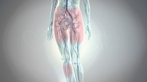
The term “syndrome” refers to a group of signs and symptoms primarily occurring together, characteristic of a specific condition. The nutcracker syndrome (in a venous or even broader scope, vascular compression syndromes) is based upon decreased space between the superior mesenteric artery and the aorta in the area of the left renal vein.
Nutcracker Syndrome
Like most of these compression syndromes in the abdomen and pelvis, this entity has been increasingly recognized and reported as the performance of axial imaging studies has increased. Discovering this type of vein compression (in the setting of abdominal or back pain for which the study is being performed) leaves the practitioner in a quandary, and often results in a referral for evaluation for nutcracker syndrome as the cause of pain.
Many of the patients referred for this problem have had pain for an extended period of time, and many have had extensive evaluation for other abdominal pathology, often culminating in the performance of other surgical procedures that proved unsuccessful in relieving their symptoms. Like many of the other vascular compression syndromes, these findings pose little to no health threat to the patient, but the functional limitations imposed by these problems are dramatic and the pain is potentially life changing.
Misdiagnosis As a Psychological Phenomenon
Many patients have been referred for psychological evaluation and have been led to believe the pain is “in their head” and not “real.” Seeing these patients in the office, physicians are “on alert” for narcotic seeking behavior, and possible malingering or Munchausen syndrome.
The other critical issue in this puzzle for the clinician is the understanding that he or she has seen numerous other patients with some form of compression of the renal vein without any symptoms at all. What is the role of the surgeon in this process, and how can we all best treat these patients with compassion and understanding, while providing therapy for those who need it and appropriate referral to other specialties for those needing this?
“PCS patients deal with many of the same issues as patients with nutcracker syndrome in terms of delays in diagnosis, referrals for psychiatric evaluation and gynecologic or other surgical procedures."

NCS & PCS Similarities
Adding to the lack of clarity regarding diagnosing nutcracker syndrome is its association with another complex issue—pelvic congestion syndrome (PCS). Like the nutcracker syndrome, there is often a long history with extensive testing and in many instances, referral for surgical procedures. PCS patients deal with many of the same issues as patients with nutcracker syndrome in terms of delays in diagnosis, referrals for psychiatric evaluation and gynecologic or other surgical procedures.
We know some of the basics about patients who present with these problems, and understanding the typical presentation and diagnostic maneuvers can be benefifical. Many of these patients are female typically in their 30s or 40s, although it is not unusual to see patients in adolescence. It is less common to see these individuals later in life.
Patients with pelvic congestion syndrome in particular tend to be parous women, and the syndrome is characterized by at least six months of persistent pain.
Many patients will have left flank and/or lower abdominal pain, and often macro- or microscopic hematuria. PCS has been associated with symptoms of dysmenorrhea, dyspareunia, and post-coital pain which can be aggravated in individuals who have vaginal or vulvar varicosities. In males with this syndrome, a left-sided varicocele can be a presentation mode, and pain in this varicocele can be seen with prolonged standing.
Some individuals will present with recurrent thigh-high varicosities following prior treatment as the initial finding. Diagnosing these problems is difficult because there are many people with these findings who are not symptomatic. Some have suggested a scoring system for making the diagnosis, but the sensitivity and specificity of these tools are unclear.[1]
Properly Diagnosing NCS & PCS
Beyond a history and physical exam to assess for these problems, testing to assess patients presenting with these problems can be extensive as there are often numerous issues that can potentially result in similar presentations— these need to be ruled out to ensure the diagnosis.
These tests can include coagulation testing, urinalysis and urine culture, urine cytology, cystoscopy, renal biopsy, CT or MR venography and ultimately, renal and IVC venography along with manometry. In most instances, it is incumbent upon the vascular specialist to ensure there is no systemic bleeding problem, no problem with the urine draining system and no intraparenchymal renal pathology.
If these are ruled out and vein compression is noted on crosssectional or axial imaging, then moving forward with invasive testing is indicated. Duplex evaluation can be helpful as well if the veins can be adequately visualized.
The diameter of the ovarian veins > 8mm will be associated with reflux in a high percentage of individuals and duplex imaging which can show this can be diagnostic for PCS, but in many instances it is really the axial imaging that shows the dilated veins in the pelvis associated with early filling of the veins on CT venography.[2]
Ultrasound is only helpful where clear cut determinations regarding the amount of ovarian vein reflux can be made, however this is difficult to do as it is not standardized, and is more frequently unobtainable. Diagnostic evaluation of nutcracker syndrome is somewhat easier with ultrasound. Angle measurements with respect to the superior mesenteric artery and aortic angulation have been defined as definitive in diagnosing nutcracker syndrome.
Here, diameter measurements in the antero-posterior plane of the vein in an unobstructed segment (as well as one in the plane of compression along with velocity measurements at these two points) will potentially allow assessment of whether nutcracker syndrome is present by assessing the ratio between the two measurements. Reportedly, ratios above five are diagnostic for nutcracker syndrome.[3]
One of the important issues regarding both of these diagnoses is that there are many more patients with anatomic findings of this entity than individuals with symptoms related to the pathology. In a study of asymptomatic women, Rozenblit et al noted 16 of 34 women with dilated ovarian veins, all of whom had reflux.[4]
Similarly, radiologic studies have documented compression of the left renal vein in asymptomatic individuals as well.[5] Beyond this, the treatment paradigm for these conditions varies tremendously and definitive conclusions regarding the necessary components of therapy to achieve complete symptom relief in all patients remains to be elucidated.
“One of the important issues regarding both of these diagnoses is that there are many more patients with anatomic findings of this entity than individuals with symptoms related to the pathology.”
Types of Treatement
Both of these entities can be treated with conservative, interventional or surgical therapies.[6-9] There are, however, rarely adequate numbers in these reports to make definitive management recommendations regarding outcomes, symptom resolution, and strategies to favor one therapy over another.
Moving from conservative or medical therapy is based upon the failure of the more conservative therapy rather than an understanding of clinical or anatomic features that would favor outcomes from one form of therapy over another.
And even in choosing surgical or interventional therapy, the type of procedures utilized, and the technique, approach and equipment vary widely. It’s patently clear that we understand little of these problems other than they are potentially interrelated, and that we have some successful methods that resolve symptoms in patients.
We clearly need better definitions of these problems, more objective measures of determining which individuals are affected by the compression and venous distension, and education for physicians regarding the presence of these entities and their relationships to the symptoms caused by these anatomic variants.
Additionally, it would be helpful to understand the ways in which differing treatments are applied, and how physicians can best determine the therapeutic approach for an individual. Finally, we need better interventional equipment that is “purpose-built” to resolve the compression and obliterate the pelvic venous distension.
Vascular surgeons have remained open to using new technologies. More importantly, they are open to using large data sets to help define problems, diagnostic testing and therapeutic modalities for uncommon problems. This has been highlighted by looking at unusual problems like renal artery aneurysm through the Vascular Low-Frequency Disease Consortium.[10]
Going From Retrospective to Prospective
While this approach is helpful from a retrospective standpoint, the fact is the issues with these two syndromes would benefit more from a prospective evaluation with interested individuals collecting baseline variables, and ultimately their approaches utilized for therapy. These are the types of innovative approaches the AVF could organize through its membership, which would create an environment of learning and knowledge regarding commonly misunderstood problems.
It would also allow treating physicians to contribute to a knowledge base which would benefit patients, physicians, and even corporate entities to gain insight into the problem and create purpose-built solutions. As we move forward in a new medical environment that will value innovative approaches and utilize information from large data sets (especially for less-frequent disease entities), the opportunity to be the leader in this type of approach would be incredibly valuable for the AVF and its membership.
Leading in innovation and gaining insight into therapy always brings value and improves quality for our patients.
References:
- Asciutto G, Asciutto KC, Mumme A, et al. Pelvic venous incompetence: Reflux patterns and treatment results. Eur J Vasc Endovasc Surg 2009; 38:381-6.
- Jeanneret C, Beier K, von Weymarn A, Traber J. Pelvic congestion syndrome and left renal compression syndrome – clinical features and therapeutic approaches. Vasa (2016) 45(4): 274-82.
- Zhang H, Zhang N, Li M, Jin W, Pan S, Wang Z, et al. Treatment of six cases of left renal nutcracker phenomenon: surgery and endografting. Chin Med J 2003;116(11):1782-4.
- Rozenblit A, Ricci Z, Tuvia J, et al. Incompetent and dilated ovarian veins: a common CT finding in asymptomatic parous women. Am J Radiol 2001; 176:119-22.
- Menard MT. Nutcracker syndrome: when should it be treated and how? Perspectives in Vascular Surgery and Endovascular Therapy.2009; 21(2): 117-24.
- Butros SR, Liu R, Oliveira GR, Ganguli S, Kalva S. Venous compression syndromes: clinical features, imaging findings and management. Br J Radiol 2013;86:20130284.
- He Y, Wu Z, Chen S, Tian L, Li D, Li M, Jin W, Zhang K. Nutcracker syndrome – how well do we know it? Urology 2014;83:12-17.
- Ahmed K, Sampath R, Khan MS. Current trends in the diagnosis and management of renal nutcracker syndrome: a review. Eur J Vasc Endovas Surg. 2006;31:410-6.
- Hulsberg PC, McLoney E, Partovi S, Davidson JC, Patel IJ. Minimally invasive treatments for venous compression syndromes. Cardiovasc Diagn Ther 2016;6(6):582-92.
- Klausner JQ, Lawrence PF, Harlander-Locke MP, Coleman DM, Stanley JC, Fujimura N. The contemporary management of renal artery aneurysms. J Vasc Surg 2015;61(4):978-84.


