
by Eric Ansart, Lympha Press President, US distribution; Donald Scott Covington, MD, FACS, CHWS, MED; Suzie Ehmann, DPT CWS CWLT CLT-LANA
There is tremendous value generated from improved communication between specialty providers and industry partners. Patients receive improved care when silos in the health-care industry are removed and caretakers can work together to create comprehensive approaches to tackle difficult diagnoses, such as lymphedema.1
Lymphedema is a clinical entity witnessed across a broad range of disease states. It presents in a variety of ways, from stereotypical arm swelling following breast cancer treatment to congenital lymphedema with onset during puberty. Individuals living with lymphedema have unmet needs that can only be addressed with a multidisciplinary team approach that includes health care providers and industry partners. When a siloed approach to care delivery prevails, best practices are rarely shared among the various disciplines to help manage the burden of this disease state. The purpose of this paper is to reflect on the three states of lymphatic treatment and the importance of taking a multidisciplinary approach to edema management.
The Starling Principle’s role in understanding Lymphedema
For nearly a century, it was commonly understood that edema was primarily a condition of venous hypertension, reflux, and an inability of the terminal capillaries to reabsorb the fluid released into the interstitial space. Known as The Starling Principle,2 the plausible concept of filtration from the arterial capillaries and sustained reabsorption into venous capillaries became embedded in the literature. Starling’s principle was taken to be a well-proven, but somewhat fossilized truth. Most physicians would learn of Starling’s principle in their first year of medical school.3
In the 1990s, the emergence of new technology to visualize the lymphatics led to an Important Review Article from C.C. Michel in Experimental Physiology, which concluded: I am sure that if Starling were alive today, he would be happy to know that his hypothesis had been so fruitful in clarifying thinking and encouraging experimentation. I believe he would readily accept that, because microvascular walls are permeable to plasma proteins, the movements of fluid through them and the hydrostatic and oncotic pressure gradients across them represent a steady-state rather than equilibrium. Having accepted that, he would then chide us for clinging so long to an outdated model of microvascular fluid exchange because it was so convenient to teach.4
In 2004, Adamson and colleagues set about revising the original Starling Principle based on new methods to visualize the lymphatics. This revised approach gives a much better understanding of the complexities of vascular edema. We now realize that all edema has a lymphatic component. This was the impetus for Levick’s published revision of the Starling Principle in 2010.⁵ It is important to note that veins still play an important role in the transference of lymphatic fluid; however, the old adage of the veins handling 90 percent of the reabsorption of interstitial edema is no longer embraced by thought leaders. The new, widely-accepted view is that lymphatics are responsible for the majority of the lymphatic uptake in the management of lymphatic overload.3
Therefore, lymphedema is added to the myriad of complex challenges faced by the vascular community. This paradigm shift further necessitates new technology to optimally assess and possibly treat lymphatic dysfunction.
Lymphedema is underdiagnosed and undertreated
Lymphedema is a poorly recognized clinical entity in the United States, with estimates suggesting only 20 percent of the patients with lymphedema are properly diagnosed and even fewer being appropriately treated.6,7
As noted previously, lymphatic overload has two primary presentations: lymphatic overload and lymphatic dysfunction. When many think of lymphedema, they consider the hereditary nature of lymphedema and reflect on the patient who either presents with chronic edema in their prepubescent period or post-cancer patients whose lymph node removal or radiation treatment has ablated their lymphatics.
At seven weeks of age, the human fetus generates lymphatic branches from the cardinal vein by forming a cluster of lymphatic sacs.8 The fundamental structure of the vascular system is established at this stage in development. Therefore, it is reasonable to consider that any malformation in the venous anatomy would carry over to the development of the lymphatic system.
As lifespan expectancy improves, it is much more common to see older patients presenting with edema. We have also recognized that hereditary lymphatic disease may remain dormant, with patients predisposed to lymphedema frequently reporting their first symptoms not until their 50s or 60s.9, 10, 11 For this patient population, the malformation of the lymphatics remains asymptomatic for decades until triggered by a traumatic event. Lymphedema is often recognized when it is the result of cancer-related interventions such as lymphadenectomy and radiation therapy. Such trauma may result from the overwhelming impact of years of untreated venous hypertension, which exposes the underlying lymphatic dysfunction and causes secondary lymphedema to become apparent.
The modern, patient-centered approach to lymphatic disease should consider these broad categories:
- Diagnosis and intervention
- Education and oversight
- Management and recovery
Lymphedema diagnosis and intervention
A basic tenant in lymphatic disease management is to recognize early symptoms and intervene appropriately. Statistics reveal that lymphatic education is grossly lacking. In a 2017 poll of major medical institutions12 it was reported that most four-year medical programs spend less than thirty minutes on the lymphatics. Once in practice, we can certainly assume that physicians in the fields of venous or lymphatic treatment are much more educated on the subject. However, a case can be made that there is a basic lack of knowledge when it comes to identifying lymphedema in general practice. The front line of care is to recognize lymphatic dysfunction in its early stages when it is more treatable and, possibly, even reversible.
Lymphedema has four stages:
Stage 0 – At this stage, edema is not always recognizable. Patients with hereditary lymphedema may remain asymptomatic in Stage 0 for decades before a triggering event causes the edema to become visually present. Bioimpedance devices like Impedimed are very effective in identifying small changes in lymph fluid collection and can help in the early identification and diagnosis of lymphedema.
Stage 1 – The development of trace structural changes and fibrotic tissue causes patients to advance in the lymphatic disease continuum. Stage 1 stems from a collection of fluid that is recognizable by the patient and identifiable by a clinician.
Stage 2 – This is a critical stage for care because the patient’s edema no longer naturally resolves with elevation. The development of fibrotic tissue has occurred and advanced therapies are required for effective treatment. Recent imaging of the lymphatics indicates that dermal backflow is evident at this stage,13 suggesting increased pressure may be needed to stimulate lymphatic return.
Stage 3 – There is often a Stage 2b that clarifies the progression between stages; however, the general progression between Stage 2 and 3 lymphedema is the significant development of fibrosis14 and hyperkeratosis (skin thickening). In Stage 3 cutaneous papilloma, papilloma and other complicating factors may develop.
The lymphatic and vascular systems stem from the same source; therefore, it is important to recognize and treat venous reflux. By doing so, symptoms associated with the edema are mitigated.
While treatment is important, chronic edema is protracted by nature. An education program is crucial for superior outcomes.
Education and oversight
Lymphedema is a progressive, chronic condition that is universally under-recognized and undertreated.15, 16 Several obstacles to care are present during the assessment, diagnosis, initial intensive treatment, and long-term management. More specifically:
- Assessment – Lack of awareness, knowledge, skilled providers
- Initial intensive treatment – Access to skilled providers. Often a diagnosis is made, but there are no referrals. Also, financial resources can be a challenge.
- Long-term management – Inability to apply compression effectively
It takes a TEAM to effectively manage lymphedema. Lymphedema Therapists play a crucial role in the management of edema. Not only do they provide education to patients, but they also provide the gold standard of lymphedema care. Complete Decongestive (Physio) Therapy or CDT is a comprehensive program for reduction of edema and lymphatic health.
CDT traditionally consists of five components: 1) skin care, 2) manual lymphatic drainage, 3) compression (compression bandages, compression garments and compression pumps), 4) remedial exercise, and 5) education regarding self-care management.
CDT has traditionally been broken down into two phases: The intensive phase and the home or maintenance phase. The intensive phase is provided in a clinic under the supervision/care of a medical provider and a specialized trained therapist. The techniques learned during the intensive phase then transition to the home or maintenance phase. The ultimate goal of CDT is that the patient learns self-care techniques to manage lymphedema and optimize quality of life.
CDT is comprised of five care processes:
Education – perhaps the most crucial aspect of the CDT program. Patients recently diagnosed with lymphedema are often overwhelmed and uncertain of process of care. A qualified therapist instructs the patient on self-care methods necessary for long-term management.
Skin Care – skin breaks can cause a lymphedema patient to incur infection. Keeping skin healthy, unbroken, and well moisturized are important preventative measures.
Exercise – even small improvements in exercise are fundamentally important. Research conducted at the University of South Florida by Joshua Scallan Ph.D. and team demonstrated the impact of lymphatic flow on the health of lymphatic valves and vessels.17 Deep breathing exercises, foot flexors, and pool programs are all excellent methods of exercise for patients with limited mobility.
Manual Lymph Drainage (MLD) – developed by Dr. Emil Vodder in the 1930s. This unique technique of stretching and massaging tissue to improve lymph uptake and transfer provides a gentle pumping action on the skin. This has been shown to increase the contraction rate of lymphangion, increased reabsorption of proteins, reduced micro-lymphatic hypertension, and improved collateral lymph drainage between the lymphatic territories of the skin.
Compression – Because a lymphedema-affected extremity is damaged, it is unable to provide sufficient resistance to the build-up of fluid in the interstitial tissue spaces. The application of external compression provides the necessary support for those tissues that lost elasticity and compensates for the elastic insufficiency by increasing the tissue pressure.
Management and recovery
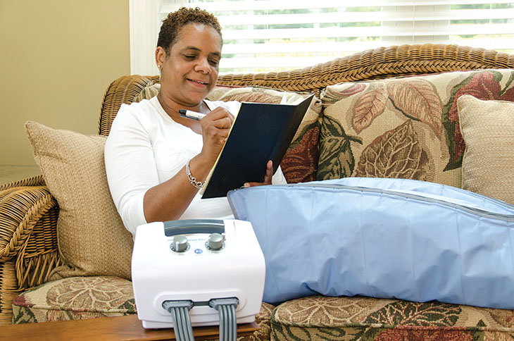
Involving industry is crucial to providing comprehensive care for the lymphedema patient population. While procedures and skilled therapy are paramount, the chronic nature of lymphedema requires that patients understand how to manage the condition at home. Proper garments, dynamic compression devices (commonly referred to as lymphedema pumps), and ancillary therapy options can all be used in this stage to improve the patient’s quality of life and outcomes.
- Garments – Compression garments can be acquired over-the-counter or by prescription. Prescribed garments tend to be of higher quality and allow for customization when needed. Robyn Bjork and Suzie Ehmann have published a comprehensive guide to selecting the proper compression garment.18 These options include:
- Flat knit – Made with a machine that knits in sheets, producing a more dense and stiff fabric with a gauge of 2 to 10.
- Circular knit – Seamless fabric with a gauge of 12 to 22 (The higher the gauge is, the thinner the fabric).
- Velcro assisted wraps – Products such as the Sigvaris Compreflex and the Farrow Wrap provide simple, effective ways for patients to apply gradient compression to their limbs with relative ease. These products have been shown to be as effective as wraps.
- Dynamic Compression (commonly referred to as compression or lymphedema pumps) – A number of device options are available to patients and improvements in technology have made these products more effective than ever. The key to selecting the best device for the patient is ensuring (1) ease of use (2) ability to modify treatment to the patient’s specific needs, and (3) ensuring distal hold sequence to reduce reflux.
Among the population of lymphedema patients, cellulitis is seventy-one times more prevalent. These reoccurring bouts of infection continue to degrade the lymphatics. Therefore, it is key to utilize tools that mitigate the impacts of lymphedema. The use of compression has been shown to decrease recurring cellulitis.19
Summary
Silos in health care are obsolescent. Interdisciplinary cooperation and integrated care delivery systems are vital to optimize patient outcomes. It is necessary to improving education to front line care providers and implement procedures to reduce the burden of edema. Patients can be offered permanent solutions that eliminate troublesome veins creating the edema. Including physical therapists trained in the management of lymphatic disorders allows for patients to obtain optimal results through skilled treatment while gaining the education necessary for long-term management. Lastly, involving industry to provide patients with tools for home treatment empowers them to take ownership of their condition. When patients are advocates and actively engaged in their treatment, they enjoy improved outcomes.
Edema and lymphedema are complicated conditions that require a multidisciplinary approach to care. In many ways, our understanding of the intricacies of the lymphatics is ongoing. As we learn more about how the lymph and venous systems interrelate, our approaches to care will continue to evolve. However, the need for an interdisciplinary approach to edema management will likely remain the gold standard to obtain optimal results.
References:
- Babiker A, El Husseini M, Al Nemri A. (2014) Health care professional development: Working as a team to improve patient care. Sudan J Paediatr: 9–16. PMCID: PMC4949805 PMID: 27493399.
- Starling, E. H. (1898). The production and absorption of lymph. Textbook of Physiology, chap. 1, ed. Schafer, E. A., 285-311.
- Levick JR. (2004).Revision of the Starling principle: new views of tissue fluid balance. J Physiol. 557 (Pt 3),704, doi:10.1113/jphysiol.2004.066118.
- MICHEL, C.C. (1997). Starling: The formulation of his hypothesis of microvascular fluid exchange and its significance after 100 years. Experimental Physiology, 82, 1-30.
- Adamson RH, Lenz JF, Zhang X, Adamson GN, Weinbaum S, Curry FE (2004). Oncotic pressures opposing filtration across non-fenestrated rat microvessels. J Physiol., 557(Pt 3),889-907.
- Rockson SG, Granger DN, Skeff KM, Chaite W. (2004).Lymphatic biology and disease: Is it being taught? Who is listening? Lymphatic Res Biol, 2,86–95.
- Korea Jong Kyoung Choi, MD, Hui Dong Kim, MD, Young Joo Sim, MD, PhD, Ghi Chan Kim, MD, PhD, Dong Kyu Kim, MD, Byeng Chul Yu, MD, PhD, Si-Sung Park, MD, PhD, and Ho Joong Jeong, MD, PhD. A Survey of the Status of Awareness of Lymphedema in Breast Cancer Patients in Busan-Gyeongnam.
- Yang Y, García-Verdugo JM, Soriano-Navarro M, Srinivasan RS, Scallan JP, Singh MK, Epstein JA, Oliver G. Lymphatic endothelial progenitors bud from the cardinal vein and intersomitic vessels in mammalian embryos.
- S. Rockson (2008). Secondary Lymphedema: Is it a Primary Disease? Lymphatic Research and Biology, doi: 10.1089/lrb.2008.6201.
- Brice G, et al. (2002). Analysis of the phenotypic abnormalities in lymphoedema-distichiasis syndrome in 74 patients with FOXC2 mutations or linkage to 16q24. J Med Genet., 39(7), 478–483. doi: 10.1136/jmg.39.7.478.
- Mortimer PS, Rockson SG. New developments in clinical aspects of lymphatic disease. J Clin Invest. 2014;124(3):915–921. doi: 10.1172/JCI71608.
- Stanley G. Rockson, MD (2017). Lymphatic Medicine: Paradoxically and Unnecessarily Ignored, Lymphat Res Biol,15(4), 315–316. Published online. doi: 10.1089/lrb.2017.29033.sr PMCID: PMC5787933 PMID: 29252138
- Hiroo Suami, Asha Heydon-White, Helen Mackie, Sharon Czerniec, Louise Koelmeyer, and John Boyages. A new indocyanine green fluorescence lymphography protocol for identification of the lymphatic drainage pathway for patients with breast cancer-related lymphedema.
- Karen Ashforth. https://lymphaticnetwork.org/news-events/understanding-lower-extremity-phlebolymphedema-fibrosis.
- Papadopoulou MC et al (2012). Multidisciplinary lymphedema treatment program. Int J Low Extreme Wounds. (1):20-7. doi: 10.1177/1534734612438436.
- Moffatt C, Keeley V, Quere I (2019). The concept of Chronic Edema-A neglected Public Health Issue and an international Response: The LIMPRINT Study. Lymphat Res Biol. 17(2):121-126. doi: 10.1089/lrb.2018.0085.
- Boksik Cha, Zeinab Y. Motawe, R. Sathish Srinivasan, Joshua P. Scallan . VE-Cadherin Is Required for Lymphatic Valve Formation and Maintenance Ying Yang, https://www.cell.com/cell-reports/fulltext/S2211-1247(19)30976-3.
- Bjork R, Ehmann S. S.T.R.I.D.E. Professional Guide to Compression Garment Selection for the Lower Extremity. J Wound Care. 2019;28(Sup6a):1‐44. doi:10.12968/jowc.2019.28.Sup6a.S1
- Kimberly M Brayton, Alan T Hirsch, Patricia J O Brien, Andrea Cheville, Pinar Karaca-Mandic, Stanley G Rockson. (2014) Lymphedema prevalence and treatment benefits in cancer: impact of a therapeutic intervention on health outcomes and costs
