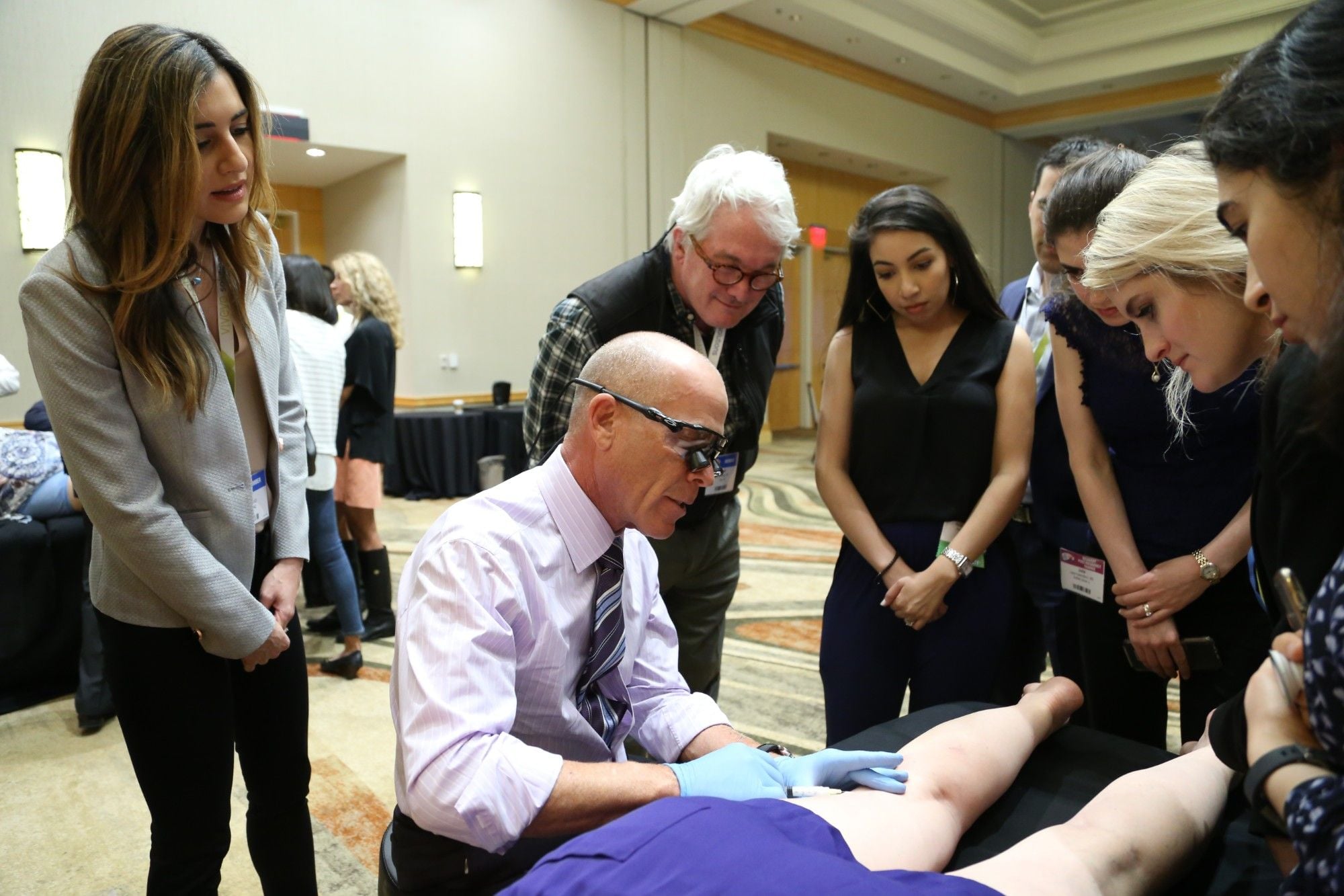
By Elika Hoss, MD and Mitchel P. Goldman, MD
Cosmetic Laser Dermatology, San Diego, CA

It is estimated that up to 20 percent of adults have varicose veins and up to 50 percent of women will develop telangictasias by the age of 50.1,2 While many patients present for cosmetic treatment, others develop adverse sequelae from lower extremity vein disease that requires medical management. When evaluating a patient with lower extremity venous disease, it is necessary to be able to identify the category of disease and choose the appropriate treatment agent and technique to maximize results and minimize complications.
To correctly perform treatment of lower extremity veins, it is first essential to understand the anatomy of the superficial vein system. The most important superficial veins in the lower extremities are the great saphenous vein (GSV) and the small saphenous vein which communicate with the deep system through perforator veins. Other superficial veins such as tributaries and collateral veins make up the venous network of the superficial system.1 Size distinguishes between varicose, reticular, and telangiectatic veins. Tortuous veins larger than 4-5 mm in diameter are referred to as varicose veins, while those between 1 and 4 mm are referred to as reticular veins, and those less than 1 mm are telangiectasias. A common myth when treating superficial leg veins is that varicose, reticular and telangiectatic veins can be treated independently of one another. But given that all superficial veins are interconnected, with blood flowing from one to the next, they are ideally treated in one procedure for the greatest efficacy.2
Sclerotherapy involves the injection of a liquid or foam substance into dilated or visible veins of the superficial system. When the sclerosing agent comes in contact with the vessel wall, a controlled thrombophlebitic reaction takes place. The term sclerotherapy comes from the Greek word “Scleros,” which translates to ‘hard’,’ describing the fibrosis that occurs due to this inflammatory reaction.3,4 Sclerotherapy is used primarily for the treatment of veins ranging between 1-4 mm, but can additionally be used for saphenous vein and perforator vein reflux. The most common sclerosing agents include hypertonic saline, chemical irritants such as chromated glycerin, and detergent solutions such as sodium morrhuate, sodium tetradecyl sulfate (STS), and polidocanol (POL). Of these, the detergent solutions can be used as both a liquid and foam, the latter of which will be focus of this discussion.1,2,4
Foam Sclerotherapy
Foam sclerotherapy involves the addition of a gas (CO2/O2, CO2, air, etc.) to a liquid sclerosing agent to create a foam.1 Initial attempts at foam sclerotherapy occurred in the 1940s, but was modernized by Tessari and colleagues in 1997, and further improved upon by Frullini in 2000. To produce foam, two syringes are connected with a three-way stopcock or a luer-luer connection. One syringe is filled with air, and the second with liquid sclerosant. The contents are then passed back and forth 10-15 times to create a uniform, white, homogenous foam. The foam must be injected quickly as it degrades one to two minutes after mixing.3,5 When injected, the foam fills the caliber of the vessel and displaces the blood, unlike a liquid sclerosant which quickly is diluted by the blood within the vein. This allows prolonged contact between the sclerosant and the vessel wall and greater endothelial cell injury. Foam is also better at producing a vasospastic response than liquid, increasing the likelihood of vein closure. Through this method, lower volumes and concentrations of sclerosant can be used effectively, while minimizing systemic and local side effects.2,4
The first European Consensus meeting on foam sclerotherapy was held in 2003, outlining guidelines and recommendations for foam sclerotherapy.6. At that time, it was determined that 6-8 mL was the maximum volume of foam sclerosant to be used per session. A second meeting in 2008 increased this maximum to 10 mL per session per leg. These restrictions are due to safety concerns for rare neurologic events or venous thromboembolism in the setting of a patent foramen ovale (PFO). The recommended ratio of sclerosant to air is 1:4 but the effect of different liquid-gas ratios on foam stability and efficacy has also been controversial.6-7
A recently completed, prospective, randomized, double-blinded, split leg study of 30 patients at our practice who were treated with standard technique found no difference in efficacy on day 21 or day 90 between two different polidocanol to air ratios, 1:2 versus 1:4. Mean improvement was between zero percent to 50 percent at day 21, and 26 percent to 75 percent at day 90 for both extremities. There was no statistically significant difference in adverse events such as erythema, swelling, urticaria, pigmentation, ankle/pedal edema, ecchymosis, ulceration, or matting between the two different polidocanol to air ratios by investigator or subject assessment. No serious adverse events were reported. Given similar safety and efficacy, the 1:4 ratio allows for treatment of a larger surface area, with a lower overall concentration of sclerosant.
A systemic review on foam sclerotherapy, estimated adverse events to occur in zero percent to 5.7 percent of cases. Minor thrombophlebitis (0.1-1.2 percent incidence), matting or skin pigmentation or hyperpigmentation (17.8 percent), and pain associated with the procedure (25.6 percent) were noted to be the most common adverse events and were generally mild and transient in nature. Other less common, minor adverse events included new vein development, nerve damage, skin necrosis, vasovagal events, chest tightness, and coughing. Serious adverse events were rare.8
In 2010, Palm et al completed a retrospective review of outcomes and adverse events of patients who had undergone foam sclerotherapy for reticular veins at one private practice center. Of the 325 patients who received foam sclerotherapy, adverse events such as hyperpigmentation, ulceration, pain, and matting or new vessel formation were reported as none or mild in severity. Average overall improvement was rated as moderate on a four-point scale. Six patients had a localized thrombophlebitis with coagulum formation requiring simple drainage. No patients reported chest tightness or pain, cough, dyspnea, visual disturbances, migraine, transient ischemic attack, stroke like symptoms, or deep vein thrombosis. One patient reported “elevated cardiac enzymes” one month after treatment, thought to be unrelated to sclerotherapy. Another patient reported “a sensation of gas leaving her lungs” in the hour after her first sclerotherapy session. A subsequent session with a similar volume of foam did not produce these symptoms. Patients received an average of 2.2 sclerotherapy sessions (range 1-2 mL), the most recent being 3.8 years prior. An average of 16.9 mL of foam were used per treatment (range 3-45 mL), which is notably higher than the consensus guidelines.3
Foam sclerotherapy has been used for decades with a high safety profile and superior results to liquid sclerotherapy, but rare serious adverse events have been reported. These events are often associated with pulmonary toxicity or more frequently an occult PFO. In the general population, more than one out of four people may have a PFO. These patients carry an increased risk for migraines, visual disturbances, stroke and cardiopulmonary events. Reports of migraines, scotomas and visual disturbances are reported in less than 2 percent of sclerotherapy patients, with later confirmed PFO in several cases.9 This is likely due to release of endothelin from the damaged endothelial cells causing vasoconstriction.10 Additionally, reports of DVT, pulmonary embolism, and stroke occurred after treatment of GSV reflux with foam sclerotherapy.11,12 However, several large studies exhibited a lack of serious adverse events with this procedure.13,15 At this time, the consensus meetings do not advocate for PFO screening in the sclerotherapy patient, perhaps due to the common incidence of PFO and common medical events in the adult population, as well as a lack of mechanistic explanation for foam directly triggering these events.7,11
Our Technique
In our practice, we perform a duplex ultrasound to evaluate for GSV reflux prior to the initiating sclerotherapy. In the case that GSV reflux is identified, sclerotherapy would be avoided until the GSV is closed by other means such as laser or radiofrequency ablation, adhesive agents, or ultrasound-guided foam sclerotherapy. If one were to proceed with sclerotherapy of the superficial venous network in the setting of GSV reflux, they would increase the risk of recurrence, hyperpigmentation, and superficial thrombophlebitis.1
Foam is generated in a 1:4 ratio, as described by Tessari.3 Injection is done proximal to distal, starting with larger reticular veins. This allows infusion of the sclerosing agent to occur through the superficial system more easily. Through this technique, one will see the solution entering the reticular vein and infusing through the venous network to the smaller distal telangiectasias, obviating the need to cannula them individually. The quantity injected should be enough to uniformly fill the vessel and displace intravascular blood, until it is no longer visualized, which means that it is flowing into the deep system.2 In our practice, we limit the amount of air to 24 mL, thus with a sclerosant to air ratio of 1:4, 30 mL of foam is safely used in one session. This is consistent with the amount of air normally released during a SCUBA dive, which has been found to be safe in millions of recreational divers over the past 50 years. After 1-4 mm veins are treated with foam sclerotherapy, the remaining smaller telangiectasias are treated with microsclerotherapy using 70 percent glycerin solution mixed 2:1 with one percent lidocaine with epinephrine 1:100,000. Immediately post procedure, patients are instructed to remain supine for five minutes to minimize blood entering the superficial venous system. Betamethasone dipropionate lotion 0.05 percent may be applied, although a randomized study by Friedmann et al, found no difference in adverse events and clearance rates compared to placebo.16
Compression therapy with thigh high class II (30-40 mmHg) compression stockings are placed and are to be worn for seven days, 24 hours a day. Compression results in direct apposition of the vessel walls, leading to more effective endosclerosis. This also limits thrombus formation, thus decreasing post-sclerotherapy pigmentation and telangiectatic matting. Walking post-procedure for 30 minutes is recommended to constrict the superficial and perforating veins and prevent deep vein thrombosis.1-4,17 Calf movements also produce rapid blood flow through the deep system to dilute any sclerosant that may have migrated to this area. Follow up is recommended at two weeks, to drain coagulums with a 22-gauge needle. Patients should be instructed that veins may look worse before they look better, due to the expected controlled inflammatory reaction. Improvement in veins will be seen for up to four months, and retreatment should not occur until at least six to eight weeks after injection to allow resolution of inflammation between treatments.2
Conclusion
Sclerotherapy is the gold standard for treatment of lower extremity reticular and telangiectatic veins. A thorough understanding of venous anatomy is necessary to perform sclerotherapy effectively. Foam sclerotherapy carries several advantages including greater efficacy, lower concentration and volume needed, and lower rates of complications such as matting or hyperpigmentation. Rare reports of neurologic and cardiopulmonary events after foam sclerotherapy exist, although true causation has not been clearly proven. With proper technique, foam sclerotherapy yields excellent results with minimal adverse events.
References:
- Goldman MP, Bergan JJ, Guex J-J. Sclerotherapy: treatment of varicose and telangiectatic leg veins. 4th ed. Philadelphia: Mosby Elsevier; 2007.
- Goldman MP. My sclerotherapy technique for telangiectasia and reticular veins. Dermatol Surg. 2010 Jun;36 Suppl 2:1040-5.
- Palm MD, Guiha IC, Goldman MP. Foam sclerotherapy for reticular veins and nontruncal varicose veins of the legs: a retrospective review of outcomes and adverse effects. Dermatol Surg. 2010 Jun;36 Suppl 2:1026-33.
- Weiss MA, Hsu JT, Neuhaus I et al. Consensus for sclerotherapy. Dermatol Surg. 2014 Dec;40(12):1309-18.
- Wollmann JCGR. The history of sclerosing foams. Dermatol Surg 2004; 30:694–703.
- Breu FX, Guggenbichler S. European consensus meeting on foam sclerotherapy, April 4-6,2003, Tegernsee, Germany. Dermatol Surg 2004; 30:709–17.
- Breu FX, Guggenbichler S, Wollmann JC. 2nd European consensus meeting on foam sclerotherapy 2006 Tegernsee, Germany. Vasa 2008;37:S1–30.
- Jia X, Mowatt G, Burr JM, et al. Systematic review of foam sclerotherapy for varicose veins. Br J Surg 2007;94:925–36.
- Ceulen RPM, Sommer A, Vernooy K. Microembolism during foam sclerotherapy of varicose veins. N Engl J Med 2008;358:1525–6.
- Frullini A1, Felice F, Burchielli S et al. High production of endothelin after foam sclerotherapy: a new pathogenetic hypothesis for neurological and visual disturbances after sclerotherapy. Phlebology 2011: 26: 203-8
- Gillet JL, Guedes JM, Guex JJ, et al. Side effects and complications of foam sclerotherapy of the great and small saphenous veins: a controlled multicentre prospective study including 1025 patients. Phlebology 2009;24:131–8.
- Bush RG, Derrick M, Manjoney D. Major neurological events
following foam sclerotherapy. Phlebology 2008;23:189–92. - Barrett JM, Allen B, Ockelford A, Goldman MP. Microfoam
ultrasound guided sclerotherapy of varicose veins in 100 legs.
Dermatol Surg 2004;30:6–12. - Hamel-Desnos C, Desnos P, Wolmann JC, et al. Evaluation of the
efficacy of polidocanol in the form of foam compared with liquid
form in sclerotherapy of the greater saphenous vein: initial results.
Dermatol Surg 2003;29:1170–5. - Guex JJ, Allarert FA, Gillet JL, Chleir F. Immediate and midterm complications of sclerotherapy: report of a prospective multicenter registry of 12,173 sclerotherapy sessions. Dermatol Surg 2005;31:123–8.
- Friedmann DP, Liolios AM, Wu DC, Goldman MP, Eimpunth S. A Randomized, Double-Blind, Placebo-Controlled Study of the Effect of a High-Potency Topical Corticosteroid After Sclerotherapy for Reticular and Telangiectatic Veins of the Lower Extremities. Dermatol Surg. 2015 Oct;41(10):1158-63.
- Nootheti PK, Cadag KM, Magpantay A, Goldman MP. Efficacy of graduated compression stockings for an additional 3 weeks after sclerotherapy treatment of reticular and telangiectatic leg veins. Dermatol Surg. 2009 Jan;35(1):53-7; discussion 57-8.


