Gray areas in vein disease: In this series of articles, we discuss the uncertain and the unsure. Eight thoughtful, knowledgeable, and confident vein specialists contemplate four venous disease areas: superficial disease; deep disease, reflux; deep disease, post-thrombotic; and acute DVT.
WE OFTEN SEE patients in our practice who have something other than the usual superficial truncal vein reflux. What should we do when patients also have an obstructed or incompetent deep vein system? There is very little data on this type of case and quite a bit of debate about how to manage these complex situations. We are forced to make challenging decisions about how to manage patients with mixed superficial and deep venous pathology. Frequently, these patients present with a more advanced stage of disease, which makes it more crucial to make the proper decisions in the appropriate order.
On the initial evaluation, any available imaging can be helpful, including CT venogram, MR venogram, duplex ultrasound of legs and abdomen/pelvis, intravascular ultrasound (IVUS), and even air plethysmography (APG), if available. After we obtain the available anatomic data, we often rely on anecdotal experience and wisdom to help make management decisions. Risk and benefit analysis must come into play. Prior attempts at treatment and their successes or failures need to be considered for each individual patient. As always, conservative measures must be maximized in these more advanced patients. The patient should be informed about their prognosis following treatment, and they should be told that the treatment offered may not fully correct their issues.
Common scenarios of deep vein pathology that can be associated with superficial vein reflux
- Chronic venous outflow obstruction proximal, abdominal/pelvic
- Iliac veins (thrombotic or non-thrombotic)
- Inferior vena cava (IVC) and iliac vein obstruction (mostly post thrombotic) – worse prognosis
- Chronic deep venous obstruction – infra-inguinal (mostly post thrombotic)
- Common femoral vein
- Femoral vein thigh
- Popliteal vein
- All segments obstructed – worse prognosis
- Deep venous insufficiency (reflux often following recanalization of deep vein thrombosis (DVT))
- Iliac/common femoral vein reflux
- Femoral vein thigh reflux
- Popliteal vein reflux
- All three segments refluxing (severe axial reflux with high maximum reflux velocity (MRV) has worse prognosis)
Case: Mr. X
A 69-year-old male with advanced chronic venous insufficiency, severe coronary disease, and thrombophilia, on long-term anticoagulation with a permanently embedded IVC filter and a chronically occluded central venous system (IVC and both iliac veins are chronically occluded). He has developed large abdominal wall collaterals and has no lower extremity venous claudication when ambulating.
He was wearing compression, which helped significantly with the edema for years, but over a few months he had gained weight and stopped wearing the compression. Since he stopped wearing his compression, he developed a small right lower extremity venous ulceration.
He is being considered for a coronary bypass with unstable angina.
Duplex exam of his right leg showed phasic flow at both groin levels and combined infra-inguinal deep and superficial vein reflux. The femoral vein had some chronic scarring but was patent and had a reflux velocity (MRV) of 10 cm/sec.1 He also had reflux in the right great saphenous vein (GSV). His right GSV measured 4–5 millimeters in diameter (an adequate size for a vein conduit).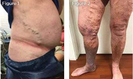
Figure 1: Patient with large abdominal wall collaterals found to have complete occlusion of inferior vena cava (IVC). Iliac veins from occluded IVC filter years ago.
Figure 2: Skin changes with new ulceration (C6) in right lower extremity after patient stopped wearing compression.
What should we do? My opinion
- He needs to maximize his conservative measures with compression, wound care, elevation, ambulation and weight loss and consider pentoxifylline or micronized purified flavonoid 3
- If the leg ulcer heals quickly with the resumption of conservative measures, the GSV should be saved for the cardiac surgeons who may need this as a conduit.
- If the leg ulcer does not heal rapidly, an ablation of the superficial vein system (GSV) could help to reduce his ambulatory venous hypertension without having a significant operative He would not tolerate a general anesthetic or a prolonged operative time if we attempted to re- establish central flow with his chronic IVC and iliac vein occlusions. He has developed enough collaterals, has phasic flow at the groin, and has no claudication in his legs. Therefore, the central venous obstruction would not be addressed at this time. His femoral vein reflux velocity is sufficiently low (MRV < 10 cm/second). Therefore, his prognosis for some relief (ulcer healing)following superficial vein ablation is reasonable.1
If he fails to heal, or if he has recurrent ulceration, we would have a risk/benefit discussion, and superficial GSV ablation would be recommended without addressing his well collateralized central venous obstruction. Although outside the area of discussion in this article, I would offer him either thermal GSV ablation or non-thermal glue (if he were not allergic) to avoid tumescent local anesthetic discomfort and risk of congestive heart failure.
In most cases, we would seek to re-establish central venous flow, but this patient has extensive collaterals, and his chronic IVC and iliac obstruction would require a high-risk procedure that he would not tolerate. Superficial vein ablation could help to lower his venous hypertension and possibly assist in ulcer healing.
Importance of collateral veins surrounding deep vein obstruction
It has been a controversial issue regarding treatment of the refluxing saphenous vein in the presence of chronic thrombotic femoral vein occlusion. Raju, et al. observed great saphenous vein flow measurements in patients with femoral vein occlusions and compared them to controls with non-occluded femoral veins. He found that there was no difference in the GSV flow with or without femoral vein occlusion. Most of the chronically occluded femoral vein patients had an enlarged profunda femoral vein acting as the deep venous outflow tract for these limbs. He found that obliteration of the great saphenous vein was successful, even in the presence of thrombotic femoral vein occlusion. He found no increase in obstructive symptoms or sequela when the GSV was closed.2
In our experience, superficial vein ablation can be done safely in most cases of chronic thrombotic deep vein occlusion if there are adequate collaterals.
We do imaging to assure the presence of an enlarged profunda femoral system of collaterals before obliterating superficial veins. In more acute settings of femoral vein occlusion, we avoid ablation of the surface veins and proceed with medical and/or procedural management to help re-establish deep vein flow if possible.
Collaterals
- Iliac obstruction can be decompressed via suprapubic or cross pelvic vein
- For femoral popliteal chronic thrombotic obstruction (CTO), there is usually efficient collateralization via the profunda femoral
- Contrary to conventional teaching, an enlarged incompetent saphenous vein system is not likely to be a significant source of collateral outflow for the legs in chronic femoral vein obstruction.2
Deep Vein Reflux and Superficial Vein Ablation
Marston, et al. have analyzed patients with mixed superficial and femoral/popliteal vein reflux and their outcomes following an endovenous ablation of the GSV (EVA). He found that there was significant improvement in venous filling index (VFI) and venous clinical severity scores (VCSS) when the deep vein reflux velocity (MRV) was less than 10 cm/sec.1 The patients who had higher femoral vein reflux velocity seemed to have less clinical improvement after EVA of the superficial system alone.
We use this MRV criteria to counsel patients on their prognosis for success following superficial vein ablation. If there is severe reflux in the femoral/popliteal veins, conservative measures are maximized. If conservative care is not successful, EVA can be performed but with less chance of full success with severe (>10 cm/sec) deep and superficial reflux. The ulcer guidelines also suggest restoration of valve function in the deep system when deep vein reflux is severe, including the following:
- Individual valve repair for those who have axial reflux with structurally preserved deep venous valves.
- Valve transposition or transplantation for those with absence of structurally preserved axial deep venous valves when competent outflow venous pathways are anatomically appropriate for surgical
- Autogenous valve substitutes by surgeons experienced in these techniques.3

References:
- Marston WA, et al. “The importance of deep venous reflux velocity as a determinant of outcome in patients with combined superficial and deep venous reflux treated with endovenous saphenous ” J Vasc Surg 2008 Aug;48(2):400-5.
- Raju S, et “Relative importance of iliac vein obstruction in patients with post-thrombotic femoral vein occlusion.” J Vasc Surg: Venous and Lym Dis 2015;3:161-7.
- O’Donnell T, et al. “Management of venous leg ulcers: clinical practice guidelines of the Society for Vascular Surgery and the American Venous ” J Vasc Surg 2014;60:3S-59S
