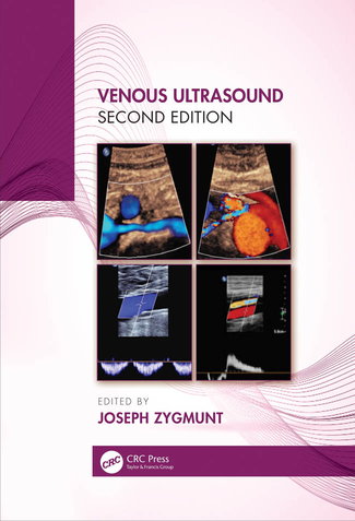
 THE FIRST EDITION of Venous Ultrasound—the definitive reference book edited by Joseph Zygmunt—was published in 2013. Back then, many physicians and sonographers were looking for a practical, step-by-step approach to saphenous duplex scanning. At the time, there was an abundance of endovenous ablations being performed, and while many providers were skilled at performing duplex ultrasounds for deep vein thrombosis (DVT), reflux testing was not as well-known or understood. The majority of venous duplex studies conducted ruled out DVT, but the DVT study did not provide adequate information for the patient with venous reflux nor for the physician performing vein ablations.
Fast-forward to 2020, and the newly-released second edition of Venous Ultrasound is here—again edited by Joseph Zygmunt and with contributions from top luminaries in the ultrasound community. Along with some updates to existing text, the newest publication features three new chapters and reflects the liveliness of the ever-evolving field of venous disease treatment. Here we highlight some (though not all) of what’s new in the second edition of this vital text.
Pelvic Insufficiency Scanning Techniques
One of the most in-demand additions that will grab readers’ interest is the chapter on pelvic insufficiency scanning techniques, as this field of interest has grown dramatically in recent years. Sara H. Skjonsberg, an accomplished sonographer who currently lectures on this topic for the American Vein and Lymphatic Soci-ety (AVLS) and their RPhS review course, authored the section. She presents this challenging information in a practical, straightforward way that allows for incorporation into daily practice immediately. For the novice in pelvic imaging, Sara describes a stepwise protocol with corresponding duplex images.
One great feature of the chapter is the inclusion of highlighted images and clear descriptions. Abnormal findings are well-shown, including tips on obtaining these images. Illustrations show pertinent anatomy with up-to-date references in this expanding field of focus.
Iliocaval Scanning and Stent Evaluation
Necessitated by the recent introduction of venous stents into the US market, the second edition also includes a new chapter on iliocaval scanning. Jan Sloves, the director of vascular imaging at Mount Sinai in New York, teamed up with Dr. Jose Almeida of Miami Vein Center to author this chapter—the two experts had previously published an article on iliac scanning in the Journal of Vascular Surgery, making them the perfect pair to pen this section.
This chapter has twenty-two pages loaded with duplex images and a panoply of tips and tricks for the sonographer looking to become adept at scanning these items. In it, Sloves provides protocols and the basics of interpretation criteria for those looking to add iliac scanning to their capabilities. Aside from criteria, the image sets—in particular, one panoramic collection of images with accompanying illustrations—provide clarity for the anatomy and challenges encountered in scanning this part of the body.
Deep Vein Obstruction
As the main focus of the first edition was saphenous scanning, deep vein obstruction scanning was missing. In this edition, Brian Fowler of Ohio Health systems added a chapter that dives into deep vein obstruction for the lower extremities and infrainguinal duplex techniques. Mr. Fowler is a key contributor to Syntropic Core Labs and is involved with new technologies and medical devices, making him a perfect choice as the author of this chapter.
Many believe the deep system anatomy to be straightforward—basically, one vein that changes name as it ascends the leg from the popliteal vein—though this is not always the case. Mr. Fowler takes us down a road of anatomic information on unitruncular and bitruncular variations. Further diagnostic criteria and protocols provided will assist anyone in preparing those documents for Intersocietal Accreditation Commission (IAC) accreditation.
Saphenous Scanning
The editor also expanded and updated the chapter on saphenous scanning, insufficiency, and reflux. In recent years, there have been many discussions at medical conferences about the overutilization of ablation procedures. An education gap exists concerning the interpretation of reflux curves, the measurement of reflux on a spectral analysis waveform, and the quality of a reflux curve, which may contribute to this issue. This edition points out “noise along the baseline,” which is called “reflux in error;” this information will help those who are misinformed on proper technique. The text shows examples of both good- and poor-quality reflux curves. After all, a low-quality study is simply non-diagnostic. Both physicians and technologists can learn from these comments.
Ultrasound Physics
Finally, one of the main updates is to the chapter on ultrasound physics. This chapter was enhanced and updated primarily for anyone serious about credentialing in venous ultrasound. Frank Miele, who many believe to be the guru when it comes to ultrasound physics, wrote this chapter. Frank is a well-known and longtime speaker at meetings, such as the Society for Vascular Ultrasound, and operates Pegasus lectures, which has other valuable materials about the field of phlebology.
Image Optimization
For those sonographers or physicians who struggle to obtain good images, Jan Sloves provides an entire chapter on optimization. Duplex equipment today has a wide range of controls that affect imaging. Black and white images can be crystal clear with relatively limited adjustments. Using color to enhance information and diagnosis takes knowledge and understanding of which controls to adjust, and Mr. Sloves leads the reader through this process with examples of images and the improved versions.
Mr. Sloves, who has garnered a reputation for image optimization techniques, generously shares his expertise with readers. The image optimization chapter will lead practitioners to night-and-day improvements in images—like trading in an old ATL duplex for a current-day Philips imager.
Rutledge and CRC press is offering VEIN Magazine readers a 20 percent discount on Venous Ultrasound and all other titles through the end of 2020. To redeem, visit https://www.routledge.com/Venous-Ultrasound/Jr/p/book/9780367354145 and enter code KML20.
The information in this article (and the textbook) are those of Mr. Zygmunt (and his contributors). This was written and submitted privately; it does not represent the opinion or position of Medtronic and is not published in his capacity as an employee of Medtronic.


