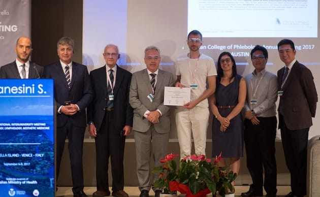
 From September 6-9, 2017, on a private island near Venice, the most renowned vein specialists gathered from around the world for the 6th International Inter-University Meeting in Phlebology, Lymphology and Aesthetics.
A faculty of more than 130 delegates came from Europe, Latin America, USA and Asia, with academics from 47 universities—all under the auspices of the Italian Ministry of Health, the International Union of Phlebology, the International Union of Angiology, the University of Ferrara and the Venous World International Network (vWIN) foundation. The related social and public venous awareness events were endorsed by the Italian Olympics Committee, the Rotary District 2060 and the Italian Golf Federation.
“From empiricism to evidence-based science” was the motto of this concept meeting, where the main focus was the analysis and divulgation of scientific data. Great attention was also dedicated to the new generations, involving them both as discussants and as speakers in a dedicated free abstract session.
Below are the five abstracts that won scientific awards out of a selection of 54.
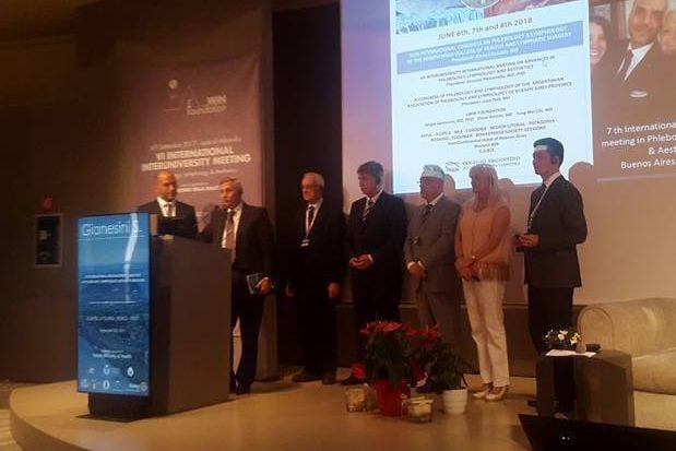
The 7th International Inter-University Meeting in Phlebology, Lymphology and Aesthetics will be held in Buenos Aires from June 6-8, 2018.
1st Oral Presentation
The relationship between disease-specific clinical and quality of life scores in chronic venous disease
Onida, S., Hameed, M., Bootun, R., Lane, T.R.A., Shepherd, A., & Davies, A.H.
Academic section of Vascular Surgery, Department of Surgery and Cancer, Imperial College London, London, United Kingdom
Background
Venous duplex ultrasound (VDUS) is the gold standard assessment tool in the investigation of chronic venous disease (CVD), providing important anatomical information to help guide treatment. CVD negatively impacts on quality of life (QoL) and correlates with disease severity. However, the relationship with the presence of reflux and vein diameter is not well-defined. The aim of this study was to compare VDUS data with disease-specific clinical and quality of life data in a population of CVD patients and healthy volunteers.
Methods
Patients with symptomatic CVD presenting in a single unit were prospectively recruited and imaged with VDUS of the lower limb(s), recording the number of trunks affected and the maximal vein diameter. The CEAP class, Venous Clinical Severity Score (VCSS) and Aberdeen Varicose Vein Questionnaire (AVVQ) results were recorded. Control subjects were similarly evaluated.
Results
Seven hundred and ninety-two participants were included: 687 symptomatic patients (51% female) with mean age 52.2 years (SD ±17: range 18-96) and 105 asymptomatic control subjects (C0-1; 62% female) with mean age 36.2 years (±12.4; range 21-88). Across the group, a weak positive correlation was identified between maximal vein diameter and AVVQ (Spearman coefficient rs= 0.276; p<0.01), CEAP (rs=0.298; p<0.01) and VCSS (rs=0.344, p< 0.01). Conversely, moderate correlation was identified between the number of trunks affected by venous reflux and AVVQ (rs=0.428; p<0.01), CEAP class (rs=0.44; p<0.01) and VCSS (rs=0.408; p<0.01).
Discussion
In a large cohort of CVD patients, a weak correlation was identified between CEAP, VCSS and AVVQ and maximal vein diameter, suggesting this may have limited utility in the assessment of the patient with CVD. Moderate correlation existed between the number of trunks affected by venous reflux and both clinical and quality of life measures, highlighting the importance of accurate VDUS assessment when assessing severity of CVD.
1st Poster Presentation
Lower limb oedema and lymphatic insufficiency following distal bypass
Oberto, S., Cetta, F., & Losa, S.
IRCCS Multimedica, Sesto San Giovanni, Milan, Italy
Background
A mainly retrospective study has been performed in 109 consecutive patients (79 males, 30 females, mean age 71 years, range 48-87) who had femoral distal bypass for critical ischemia in order to evaluate the prevalence of postoperative lower limb oedema, its evolution and likely pathogenesis.
Methods
Ninety-four patients (86%) were diabetic and only 17 patients (15.6%) had ischemic lesions classified as Rutherford 4, whereas 92 patients (84.6%) were Rutherford class 5 and 6, respectively. Thirty-five patients (32.1%) had femoral-popliteal bypass above the knee and 74 (67.9%) below the knee. Forty-three of the latter had distal anastomosis on the popliteal artery and 27 on the tibial vessels. Twenty-three had autologous saphenous vein bypass graft (BPG), 82 had heparin bonded expanded polytetrafluoroethylene (HePTFE) and 86 had heterologous BPG. Diagnosis was performed by visual inspection and triple measurement to the lower limb circumference at pre-established sites, and with Echo-Color-Duplex scanner, which led to the exclusion from the final series of subjects with concomitant or previous deep vein thrombosis. All patients were followed at 1, 3, 6 and 12 months, and every 6 months thereafter.
Results
Twenty-five subjects (22.9%) had lower limb oedema, which was moderate < 2 cm or severe > 2 cm. Twenty-three of them had complete disappearance of limb swelling after 13 to 120 days. Two patients had chronic oedema: the former underwent major leg amputation after 12 months, the latter had persistent oedema after 36 months.
Discussion
In the vast majority of patients, postoperative oedema is reversible. Excluding patients with deep vein thrombosis, lymphatic alteration or insufficiency is likely present in most of them, even if its exact role remains to be elucidated and additional factors could also play a role.
2nd Oral Presentation
Thrombophilia in non-thrombotic chronic venous disease of the lower limb — A systematic review
Hau, K. & Tan, M.
Imperial College School of Medicine, Imperial College London, London, United Kingdom
Onida, S., Lane, T., & Davies, A.H.
Academic Section of Vascular Surgery, Division of Surgery, Department of Surgery & Cancer, Imperial College London, London, United Kingdom
Introduction
Chronic venous disease (CVD) represents a significant health care burden, presenting with a spectrum of clinical signs including varicose veins and venous ulceration. Studies have proposed thrombophilia as a risk factor, particularly with respect to post-thrombotic CVD. Its relationship to non-thrombotic CVD has not been comprehensively reviewed.
Methods
PubMed and EMBASE databases were systematically searched from 1946 until March 2017. Case-control studies, cohort studies or randomized clinical trials reporting on the relationship of thrombophilia to non-thrombotic lower limb CVD in adult patients were included. Non-English and post-thrombotic syndrome studies were excluded. Study selection and data extraction were performed by two independent reviewers.
Results
Fifteen studies met the eligibility criteria, reporting on 911 cases and 1,261 controls. Studies largely focused on venous ulceration and investigated multiple haemostatic factors. A direct relationship between thrombophilia and non-thrombotic CVD was identified, with greater prevalence and factor concentration alteration reported in patients compared to controls. Presence of multiple concomitant thrombophilia was also associated with earlier CVD onset. Targeting hyperhomocysteinemia with folic acid and factor VIII, PAI-1 and von Willebrand factor with aspirin shows promise in enhancing CVD treatment. Relationship strength between each thrombophilia and non-thrombotic CVD varied, with common aetiologies like factor V Leiden and elevated factor VIII showing clearer correlation than rarer ones such as antithrombin deficiency.
Conclusion
Thrombophilia is associated with non-thrombotic CVD but causation cannot be determined. Future research should focus on prospective studies with larger study populations to establish causation and then identify adjunct therapies targeting thrombophilia.
3rd Oral Presentation
The mechanism and an incidence of antegrade diastolic blood flow in Giacomini's vein
Rosukhovskiy D.A.
FASI “Institute of Experimental Medicine”, St. Petersburg, Russia
«Dolgoletiye» Clinic, St. Petersburg, Russia
«Akson» Clinic, Viborg, Russia
Shaydakov E.V.
FASI “Institute of Experimental Medicine”, St. Petersburg, Russia
Background
Antegrade diastolic blood flow (ADBF) is described as a pathological pattern of blood flow in the Giacomini's vein (GV), associated with varicose transformation and classical reflux in great saphenous vein and tributaries. There are two theories of its appearance: the theory of “siphon”, which gives the main role to negative pressure gradient in varicose tributaries; and “overloading” theory, which means hypervolemia of GV due to blood overloading through the insufficient saphenopopliteal junction (SPJ). “Overloading” mechanism wasn’t described obviously. However, apparently, it was implied by the phlebologists, who recommended high ligation or equally obliteration of SPJ for radical treatment of varicosities associated with ADBF in GV.
Methods
Five hundred and fourteen legs of the patients, sequentially referred for the operation in 2016, were examined with duplex ultrasound. Four hundred and ninety-six legs with the recognized pattern of reflux were included in our study. We assessed the presence of ADBF in GV, GV reflux, SPJ sufficiency, SSV & GSV sufficiency.
Results
SPJ insufficiency was found in 101 (20.4%) legs and SSV reflux was found in 88 (17.8%). The GV reflux was identified in eight legs (1.6%). ADBF in GV was found in 13 (2.6%), of which three had varicose tributaries located exceptionally above the level of the saphenopopliteal junction.
Conclusions
Blood flow disturbances of the GV with clinical manifestations was present in 4.2 percent of legs with varicose veins (C2-C6, CEAP). The ADBF through the GV is observed in 62 percent of them. ADBF was strongly associated with SPJ incompetence. The location of varicose tributaries above the "escape point" of reflux (in some cases of ADBF) demonstrates the failure of the “siphon” theory.
4th Oral Presentation
Biomatrix sclerofoam: Equal or better than thermo-occlusion?
Despa, O.R., Stoyanova, K., El-Chamali, S., Brüggemann, U., Gzelachowski, S., Ragg, D.C., & Ragg J.C.
Angioclinic® Vein Centers, Interventional Phlebology, Berlin, Munich, Zurich, Germany, Switzerland
Background
Common sclerofoams (Cabrera type, including Varithena/BTG) are inferior to thermo-occlusion in regard to primary and long-term results. Such foams are of low viscosity and stability. They will collapse within 60 to 240 seconds and rapidly lose effect. With increasing vein diameters, common foams will less displace blood but float on it with zones of uncertain effects. A novel viscous microfoam, using a biomatrix based on denatured autologous blood proteins with an in vitro half-life of > 60 minutes and fast disintegration within flowing blood, was evaluated in the GSV using a safety setting to prevent foam migration via the junction.
Methods
One hundred and twenty patients (78 females, 42 males, all aged 32–81 years old) with GSV insufficiency and diameters 6–24 mm (mean: 10.3 mm), were randomized to two modalities of treatment: A) junction closure by endovenous laser in coaxial perivenous anesthesia (EVL 810 nm, ball tip or 1470 nm, slim/radial, segment length 3–20 cm), combined with catheter sclerofoam (biomatrix sclerofoam, BSF, 1% Aethoxysklerol, segment length 28–35 cm, n = 60), for the segment below; or B) EVL alone for comparison, treated segment length 38–55 cm (n = 60). Both modalities used the same PTFE vein catheter (PhleboCath 2.3 mm, OTW). BSF was deployed during catheter withdrawal. Postinterventional examinations with ultrasound were performed after two weeks and 2, 6, 12 months by independent investigators.
Results
Initial vein occlusion was obtained in all cases (120/120). There were no adverse events, in particular no thoracic or cerebral symptoms in the group receiving BSF. The patterns of echogenicity were similar in both groups at all presentations. Vein diameter regression and mild postinterventional symptoms were similar for EVL and BSF. Concerning group A, the investigators were not able to discriminate any borderline of the methods. During one-year follow-up, junction segments showed reperfusion in 2/60 cases (A, 3.33%) and 1/60 (B, 1.6%), respectively. The thigh-to-knee segments, representing the closure quality of each method, showed partial reperfusion in 5/60 cases (8.33%) after BSF and in 6/60 cases (10%) after EVL.
Conclusions
The use of BSF in the GSV, apart from the femoral junction, is safe and effective. According to ultrasound patterns and clinical follow-up, there is no significant inferiority to endovenous lasers. However, BSF is more convenient for patient and physician as no tumescent anesthesia is required. Pilot studies using BSF also in the SFJ—the SSV including SPJ, perforator veins and recurrent varicosities—will be concluded June 2017.
